Figure 6. Immunohistochemical localization of AQP1 and AQP5 in the cornea after blast exposure (B–D) compared to control (A–C). The extent of AQP1 immunostaining increased across the corneal epithelial cells of blast-exposed rabbits (B) compared to those of control rabbits (A). AQP5 immunostaining changed from a mixed membrane and cytoplasmic expression in the corneal epithelium of control rabbits
(C) to predominantly cytoplasmic expression in the basally located cornea epithelial cells after blast exposure (D). The arrows indicate plasma membrane distribution of the corneal epithelium. The image was captured with 100X (bards=100
µm) and 200X magnification (bards=50 µm).
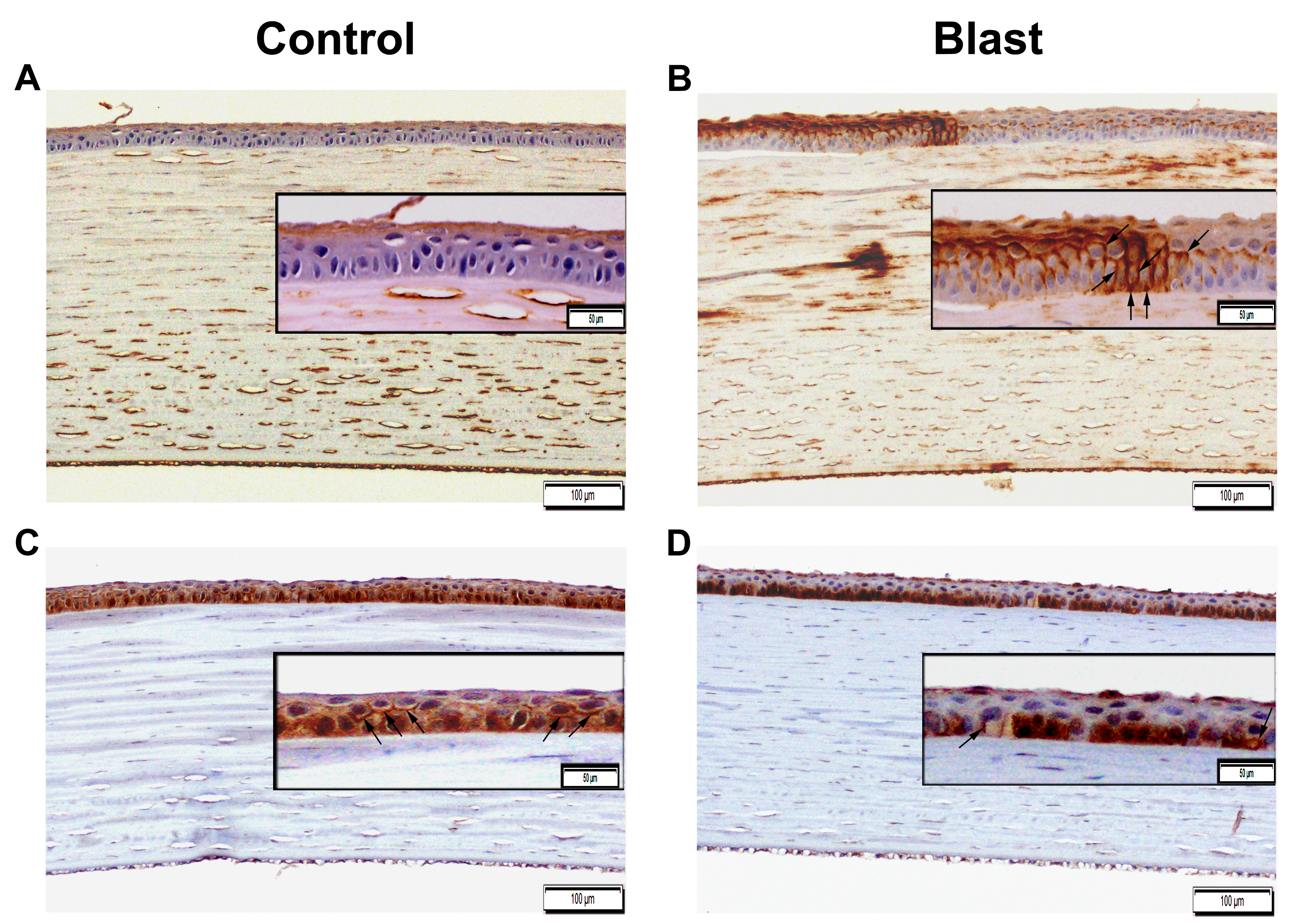
 Figure 6 of
Ríos, Mol Vis 2019; 25:283-294.
Figure 6 of
Ríos, Mol Vis 2019; 25:283-294.