Figure 5. Western blots of AQP1 (A) and AQP5 (B) proteins isolated from whole cornea homogenates. The densitometry scans of gels are shown in the bottom section of each
panel. AQP1 and AQP5 levels were significantly increased in the corneas of blast-exposed rabbits. β-actin was used as the
loading control. Arrows indicate molecular weight for AQP1, AQP5, and β-actin. Data are presented as mean ± SEM of six animals
per group.
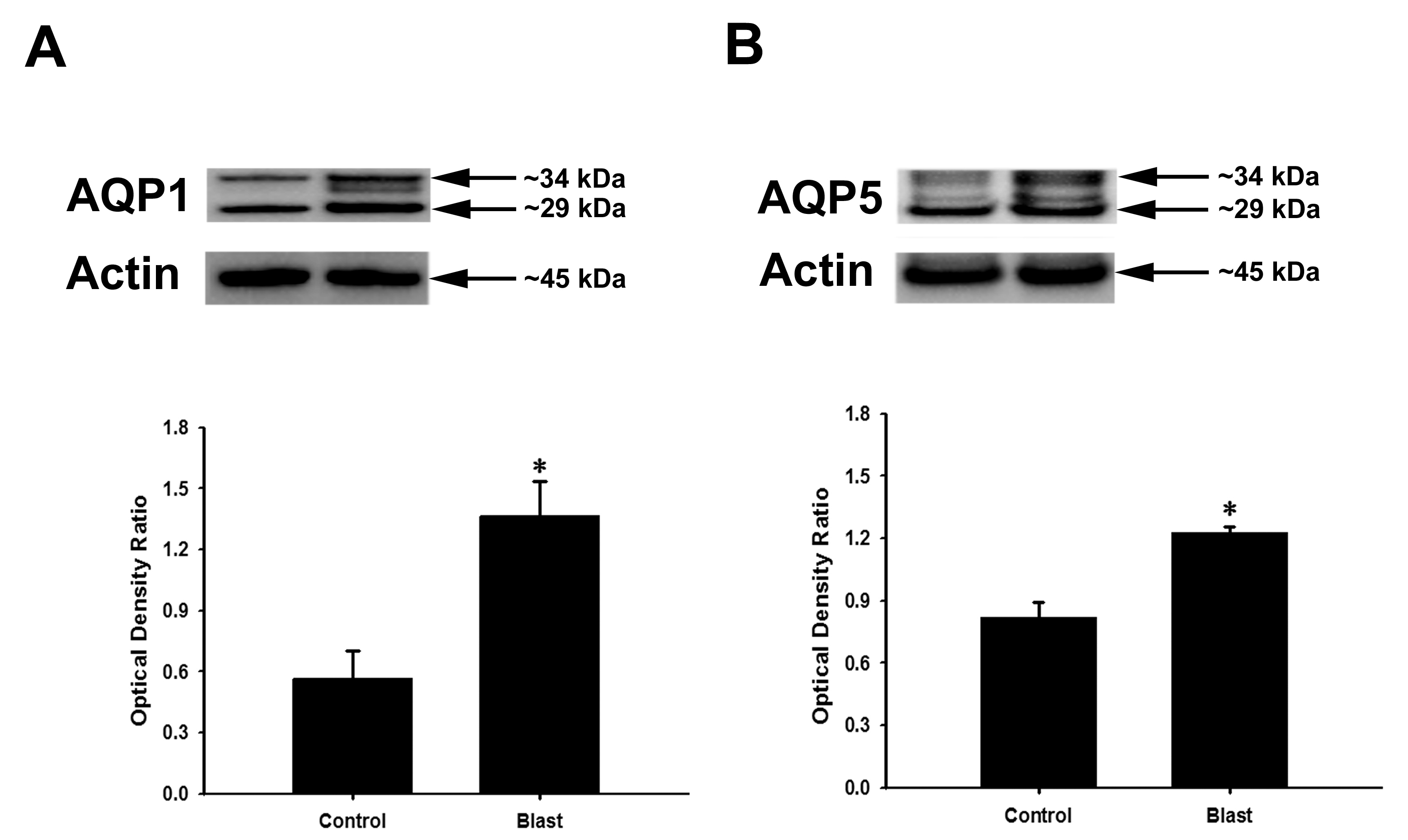
 Figure 5 of
Ríos, Mol Vis 2019; 25:283-294.
Figure 5 of
Ríos, Mol Vis 2019; 25:283-294.