Figure 6. Porcine anterior segment perfusion culture. Outflow rates were measured in porcine anterior segments perfused with 100 ng/ml
(A) interleukin-20 (IL-20) , (B) IL-19, or (C) IL-24. Cytokines were added at time point 0, and flow rates were measured for a further 73 h. The data show the average
± standard error of the mean. * p<0.001, ** p<0.01 and *** p<0.05 with ANOVA.
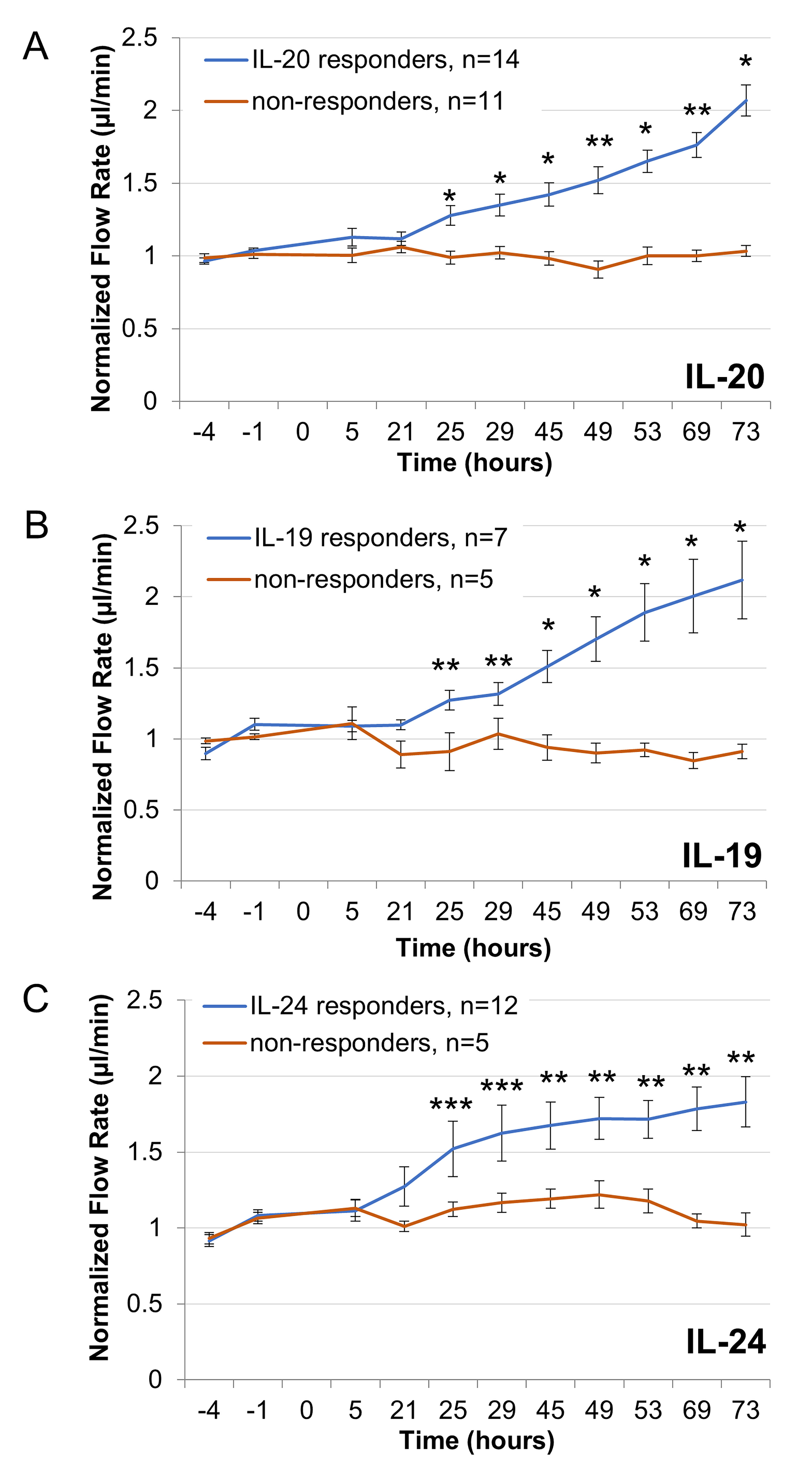
 Figure 6 of
Keller, Mol Vis 2019; 25:266-282.
Figure 6 of
Keller, Mol Vis 2019; 25:266-282.