Figure 3. IL-20R in situ protein interactions. Duolink proximity ligation assay confocal images of normal human dermal fibroblasts (A, B) or human trabecular meshwork (TM) cells (C,D). Representative images of type I (interleukin-20RA (IL-20RA)–IL-20RB) and type II (IL-22RA1–IL-20RB) receptor complexes
are shown. Scale bar = 20 µm. E: The number of spots and nuclei was counted with Imaris software in seven to 14 fields for each receptor type in at least
four different experiments using three biologic replicates for each cell type. * p≤0.024 and ** p<0.003 with one-way ANOVA.
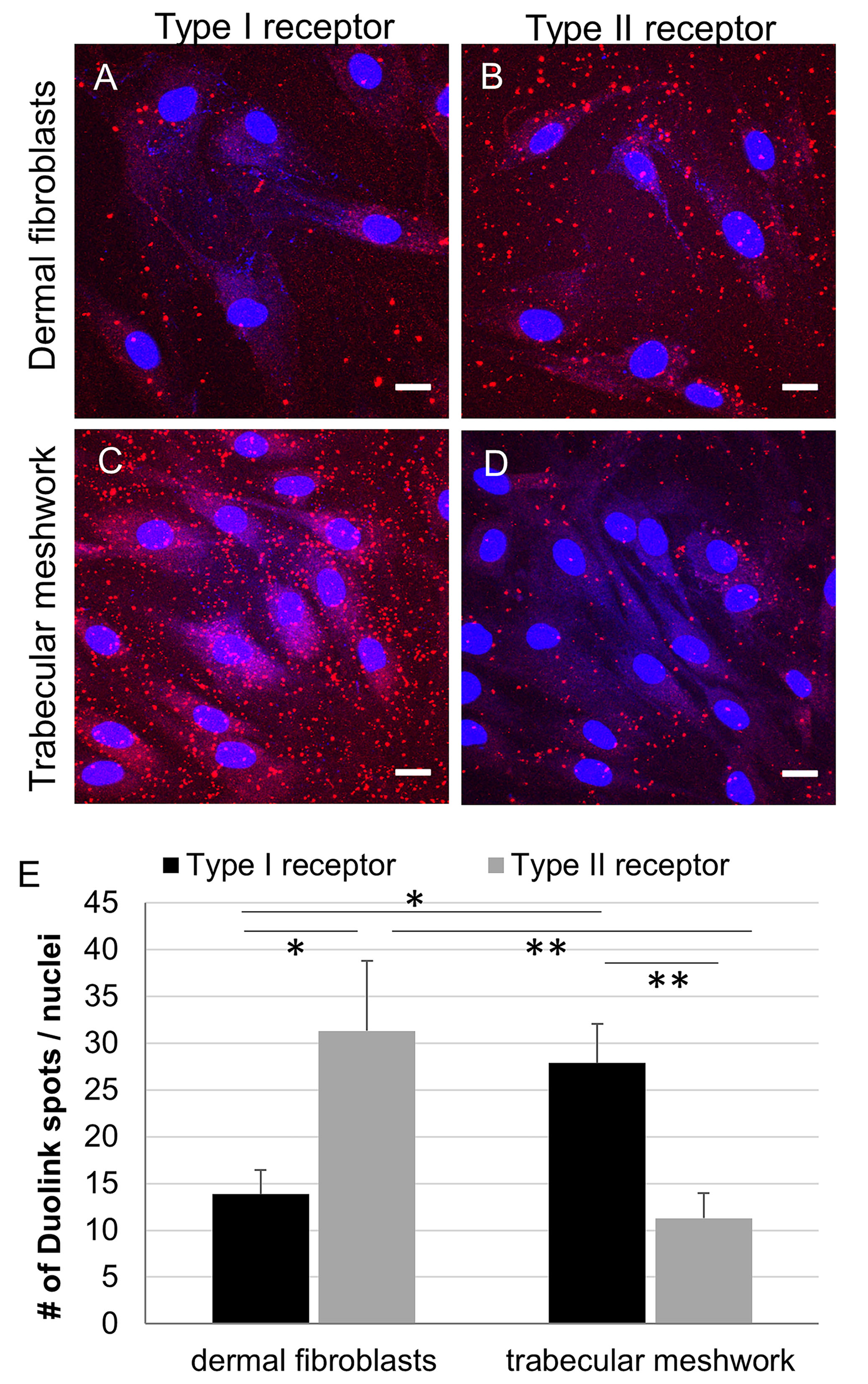
 Figure 3 of
Keller, Mol Vis 2019; 25:266-282.
Figure 3 of
Keller, Mol Vis 2019; 25:266-282.