Figure 1. Morphological and functional characterization of normal human TM cells from donors. A: A spindle-like morphology is shown in the isolated trabecular meshwork (TM) cells from donor 1 through donor 3. B: Positive staining of biomarkers laminin alpha 4 (LAMA4; red) and tissue inhibitor of metalloproteinase 3 (TIMP3; red) for
TM cells at passage 5. Cell nuclei were labeled in 4′,6-diamidino-2-phenylindole (DAPI, blue). Bar = 50 µm. C: Immunoreactive bands of myocilin proteins were visualized. GAPDH was used as the reference protein. D: The intensity of the visualized bands in the control and dexamethasone (DEX)-treated cells from each donor. Data were quantified
from three independent experiments (n=3). * p≤0.05, compared with control.
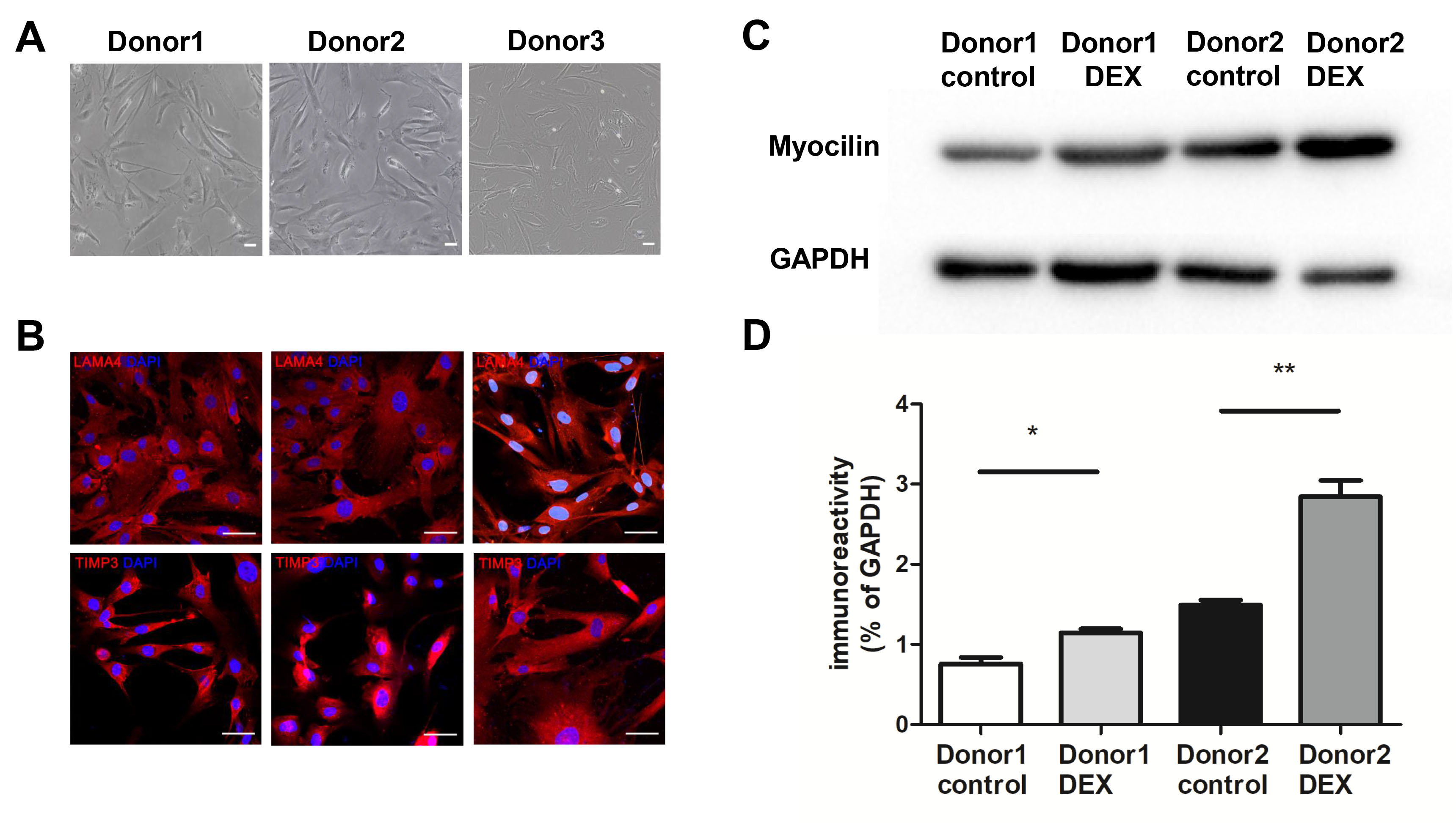
 Figure 1 of
Yu, Mol Vis 2019; 25:255-265.
Figure 1 of
Yu, Mol Vis 2019; 25:255-265.