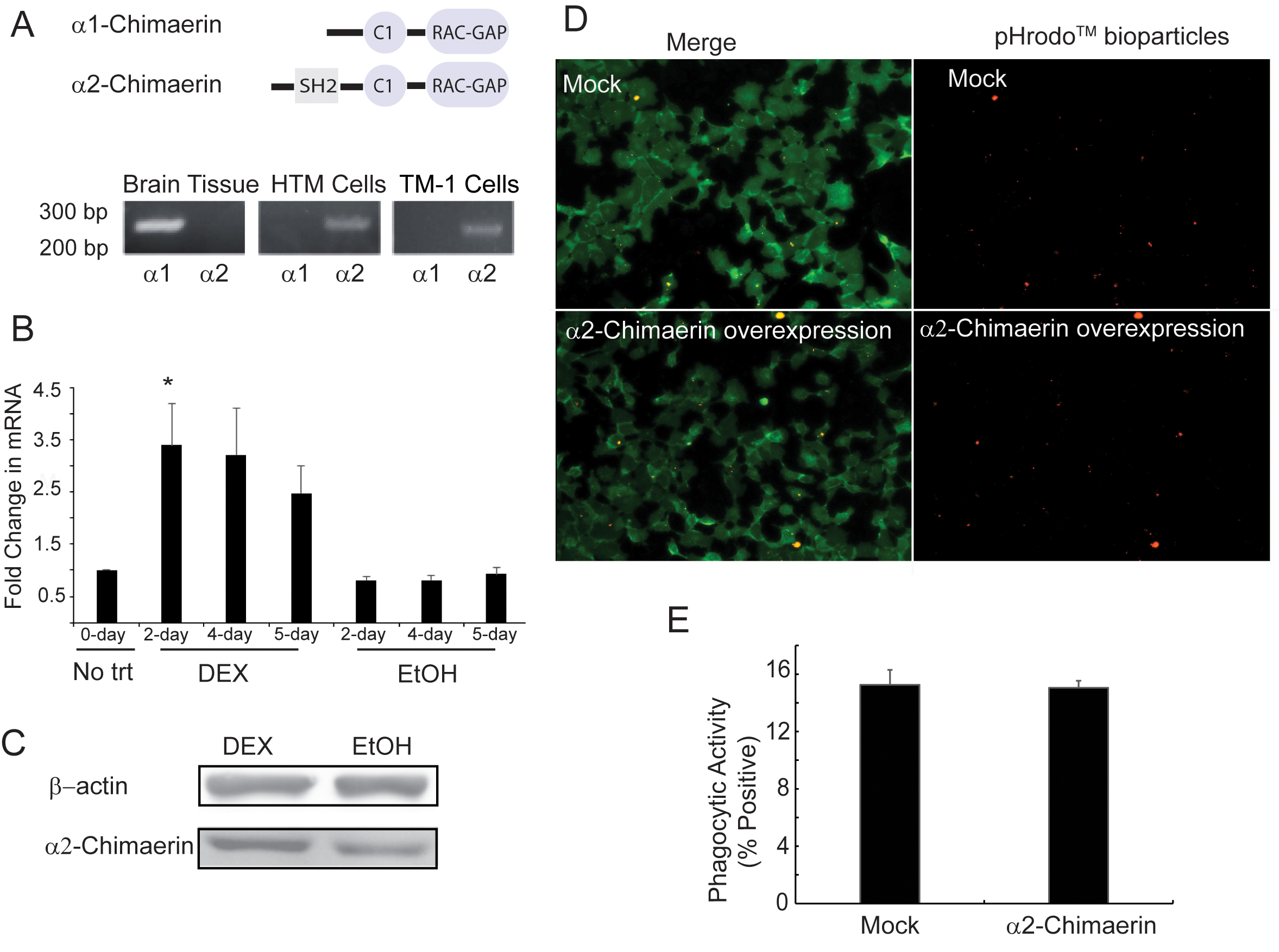
Figure 2. Expression of the RAC1-specific GAP
CHN1 is upregulated by DEX.
A: Schematic diagram showing the two splice variants of α-chimaerin. Both variants contain a GAP domain that is specific for
RAC1 and a C1 domain that mediates binding to diacylycerol and phorbol esters. The α2-chimaerin also contains a SRC homology
2 domain (SH2). PCR showed that both HTM and TM-1 cells express mRNA for the α2-chimaerin variant. In contrast, brain tissue
expressed mRNA for the α1-chimaerin variant. Primers used to distinguish between the variants were previously described [
57]. The forward and reverse primers, respectively, used to detect the α1-chimaerin variant were 5′- AAA ATG CCA TCC AAA GAG
TCT-3′ and 5′- GAA ATT GTG AAT CTT TTC ATA TTT-3′. The forward and reverse primers, respectively, used to detect the α2-chimaerin
variant were 5′-GGC TCT ACT ACG ATG GCA AGC-3′ and 5′- CTG TAG AAT CTC TCT CAT CAT GT-3′.
B: qPCR analyses showed that DEX caused the upregulation of
CHN1 mRNA compared to the EtOH-treated and no trt controls. The increase at day 2 was statistically significant (*) at p<0.05
compared to the no trt controls. Data are presented as the mean ± SEM. The mRNA levels were normalized to the no trt group.
All five HTM cell strains (N27TM-2, N27TM-4, N27TM-5, N27TM-6, and N25TM-8) were used for the qPCR analyses; n=5.
C: Western blot analyses showed that an increase in α-chimaerin levels was detected in the DEX treated cells. β-actin was used
as a loading control. Equal amounts of protein from the DEX- and EtOH-treated cell lysates were loaded. The figure is a representative
blot done on cell lysates from three separate experiments using the HTM cell strain (N25TM-8); n=3.
D: Fluorescence micrographs showed the phagocytosis of pHrodo™ bioparticles (red) in TM-1 cells overexpressing α2-chimaerin
was similar to mock transfected cells. Cells were counterstained with CellMask Green to visualize the cells.
E: Quantification of the number of cells that phagocytosed pHrodo™ bioparticles in cultures transfected with FLAG tagged-α2-chimaerin
(n=4216 cells) did not show a statistically significant change (p<0.8) in phagocytosis compared to mock transfected cells
(n=1279 cells). Data are presented as the percent positive ± SEM.
 Figure 2 of
Faralli, Mol Vis 2019; 25:237-254.
Figure 2 of
Faralli, Mol Vis 2019; 25:237-254.