Figure 4. Surgical injury induces fibrosis- and proliferation-related genes through FGF2 in the mouse corneal endothelium in vivo. A: Surgical injury induced FGF2 and endothelial to mesenchymal transition markers Snai1 and Zeb1 in the mouse corneal endothelium in vivo. This could be inhibited by fibroblast growth factor 2 (FGF2) knockdown using FGF2
siRNA. Non-targeting siRNA (NT) had no effect on injury-induced gene expression. The uninjured control corneal endothelium
showed no expression of Snai1 and Zeb1. FGF2 one-way ANOVA, F(3,8)=127.7, p=0.001, n=3 per sample. Tukey’s post-hoc test, honestly significant difference (HSD)[0.05]=1.4 and HSD[0.01]=2.0.
Snai1 one-way ANOVA, F(3,8)=18.0, p=0.01, n=3 per sample. Tukey’s post-hoc test, HSD[0.05]=1.6 and HSD[0.01]=2.2. Zeb1 one-way ANOVA, F(3,8)=25.4, p=0.01, n=3 per sample. Tukey’s post-hoc test, HSD[0.05]=3.0 and HSD[0.01]=4.2. B: Surgical injury induced expression of Col1a1, Col1a2, Fn1, and Vim, and decreased expression of Cdh1 in the mouse corneal endothelium in vivo. This could be inhibited by FGF2 knockdown using FGF2 siRNA. Non-targeting siRNA
(NT) had no effect on injury-induced gene expression. The uninjured control corneal endothelium showed no expression of Col1a1, Col1a2, Fn1, and Vim, and showed robust expression of Cdh1. Col1a1 one-way ANOVA, F(3,8)=16.3, p=0.02, n=3 per sample. Tukey’s post-hoc test, HSD[0.05]=3.5 and HSD[0.01]=4.8. Col1a2 one-way ANOVA, F(3,8)=54.9, p=0.005, n=3 per sample. Tukey’s post-hoc test, HSD[0.05]=2.6 and HSD[0.01]=3.6. Fn1 one-way ANOVA, F(3,8)=14.9, p=0.03, n=3 per sample. Tukey’s post-hoc test, HSD[0.05]=4.6 and HSD[0.01]=6.4. Vim one-way ANOVA, F(3,8)=77.8, p=0.003, n=3 per sample. Tukey’s post-hoc test, HSD[0.05]=2.3 and HSD[0.01]=3.2. Cdh1 one-way ANOVA, F(3,8)=359.2, p=0.0001, n=3 per sample. Tukey’s post-hoc test, HSD[0.05]=0.080 and HSD[0.01]=0.12. C: Surgical injury-induced expression of Cdk2 and Ccne1 in the mouse corneal endothelium in vivo. This could be inhibited by FGF2 knockdown using FGF2 siRNA. Non-targeting siRNA
(NT) had no effect on injury-induced gene expression. The uninjured control corneal endothelium showed no expression of Cdk2 and Ccne1. Cdk2 one-way ANOVA, F(3,8)=102.3, p=0.002, n=3 per sample. Tukey’s post-hoc test, HSD[0.05]=1.7 and HSD[0.01]=2.4. Ccne1 one-way ANOVA, F(3,8)=21.7, p=0.02, n=3 per sample. Tukey’s post-hoc test, HSD[0.05]=2.9 and HSD[0.01]=4.0. D) Keratocan (Ktcn) was used as the control for contamination of the stromal keratocytes. Col8a2 and β-actin (Actb) were used as the corneal endothelial cell (CEC) marker and the loading control, respectively. The data shown are representative
of the results in three independent experiments. C, Non-injured normal corneal endothelium; NT, non-targeting control. * p<0.05.
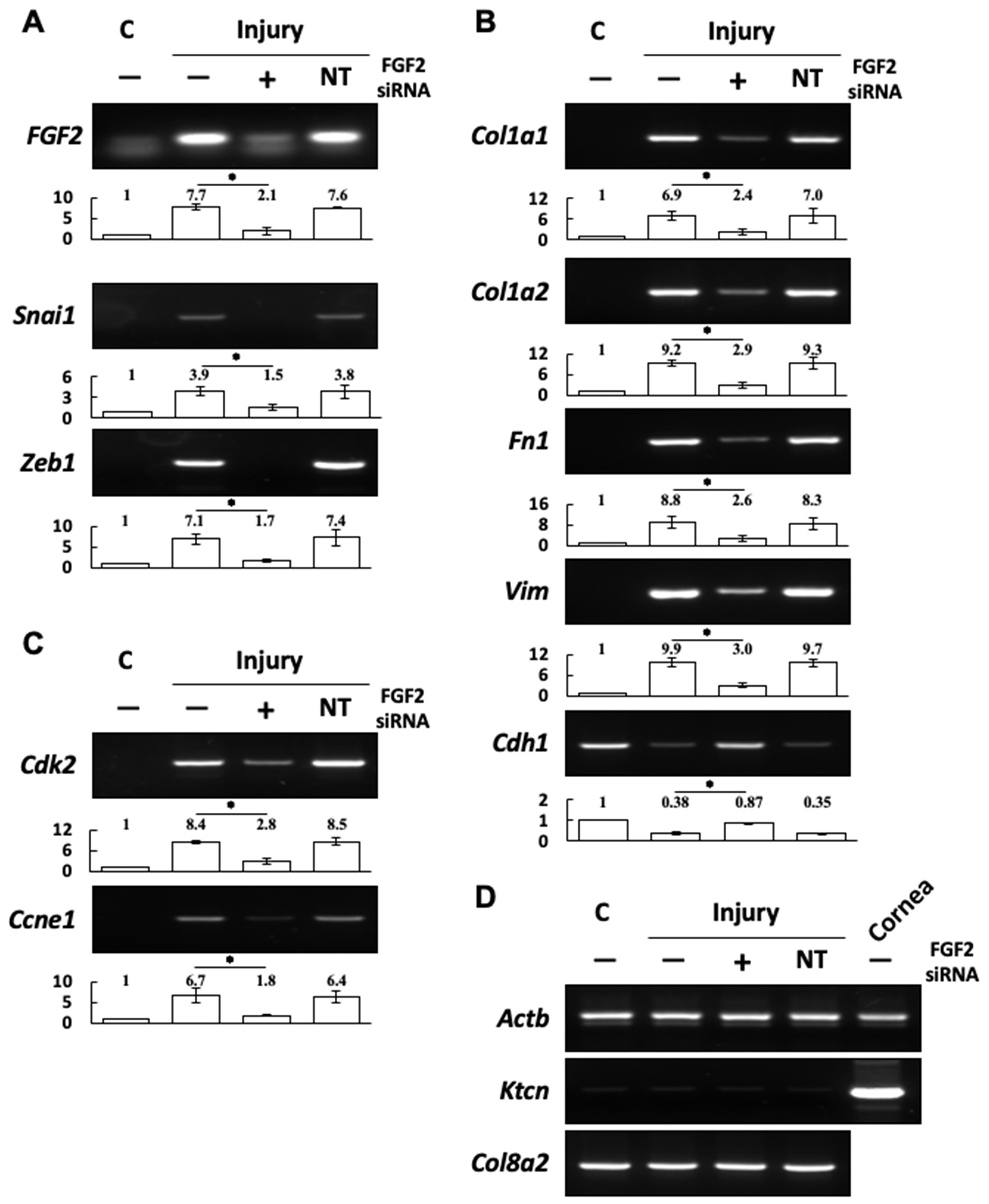
 Figure 4 of
Lee, Mol Vis 2019; 25:22-34.
Figure 4 of
Lee, Mol Vis 2019; 25:22-34.