Figure 1. Surgical injury of the corneal endothelium induces PMN infiltration into the corneal endothelium and secretion of IL-1β and
FGF2 into aqueous humor. A: At 4 weeks after surgical injury, hematoxylin and eosin (H&E) staining showed thickening and disorganization of Descemet’s
membrane with altered endothelial cells in the injured cornea. The uninjured control cornea showed a well-organized monolayer
of corneal endothelial cells. Scale bar, 50 µm. B: At 24 and 30 h after surgical injury, aqueous humor from ten anesthetized mice was collected, and immunoblotting was performed
with an anti-interleukin 1 beta (anti-IL-1β) antibody. IL-1β was not detected in the aqueous humor of the uninjured eyes,
whereas IL-1β could be detected at 24 h but not 30 h after surgical injury. C: At 24 and 30 h after the surgical injury, the aqueous humor from ten anesthetized mice was collected, and immunoblotting
was performed with anti-FGF2 antibody. FGF2 was not detected in the aqueous humor of the uninjured eyes, while FGF2 could
be detected at 24 h and increased through 30 h after injury. Equal amounts of protein (2.5 µg) of aqueous humor from uninjured
and injured mice were loaded for the immunoblotting. The data shown are representative of the results in three independent
experiments.
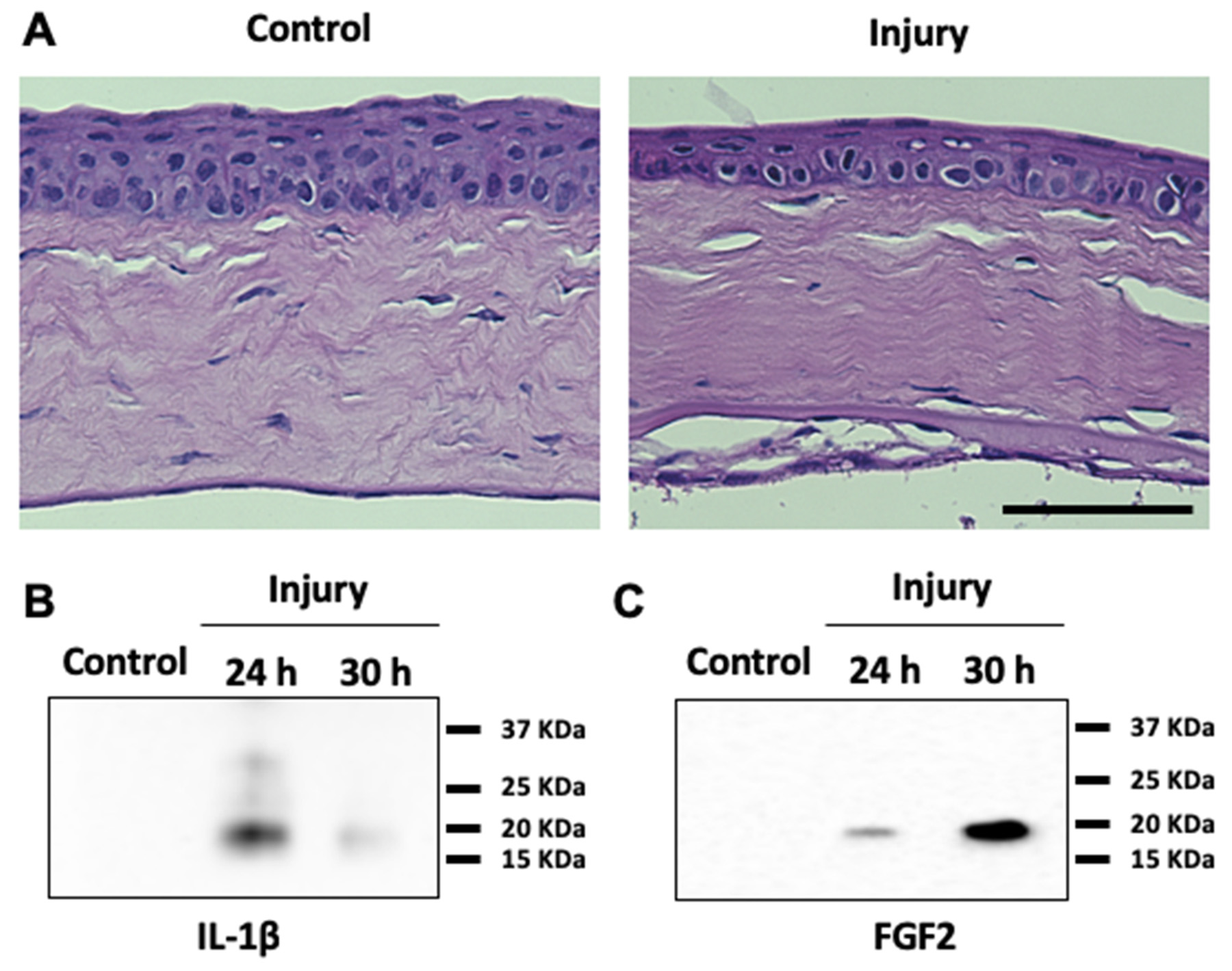
 Figure 1 of
Lee, Mol Vis 2019; 25:22-34.
Figure 1 of
Lee, Mol Vis 2019; 25:22-34.