Figure 4. Fundus photograph and optical coherence tomography pictures of three patients with mutations in RPE65. A: Patient IV:1, a 23-year-old man in Family 20061, shows disc pallor, attenuated vessels with a few early alterations in the
macula, and pepper fundus with peripheral RPE mottling. B: Patient IV:2 of Family 20061, a 32-year-old woman with a c.493C>T (p. Gln165X) mutation in RPE65 (Family 20061), shows disc pallor, attenuated vessels, scar macula, with salt and pepper fundus. C: Patient II:2 of Family 20041, a 33-year-old man with two mutations in RPE65 (p.Arg91Gln and p.Leu395SerfsX4), shows bone spicule-like formation in the fundus, attenuation of the retinal arterioles,
and optic disc pallor. D–F: Optical coherence tomography demonstrates reduced retinal thickness in these three patients with LCA.
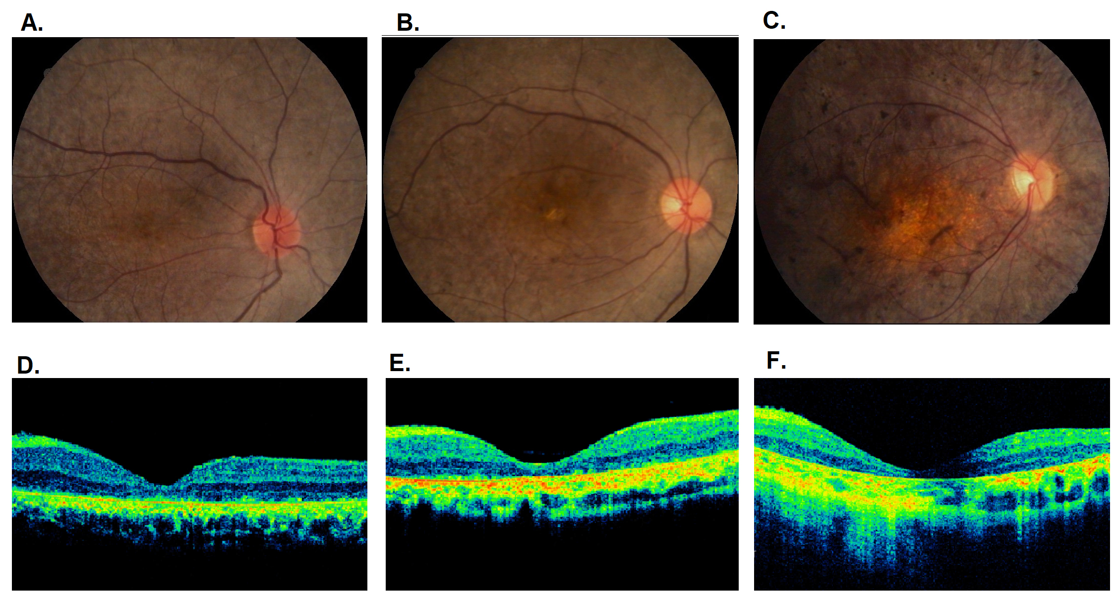
 Figure 4 of
Zhong, Mol Vis 2019; 25:204-214.
Figure 4 of
Zhong, Mol Vis 2019; 25:204-214.