Figure 3. Splicing assay shows mutation-induced change in RPE65 splicing. Gel electrophoresis of reverse transcriptase (RT)–PCR products for all tested constructs. The control is unmodified
pCAS2 (a generous gift from Dr. A. Martins, University of Rouen, France). The differences between the respective wild-type
(wt) and variation (var) band composition demonstrate the mutation-induced aberrant at the mRNA level. Dark exons: the first
and last exon of the pCAS2 reporter minigene, white exons: the exons cloned in for RPE65 mutation testing. We sequenced the band to verify the sequence of the splicing products with Sanger sequencing. A: Minigene-splicing assay of the c.858+1delG variant in RPE65. B: Minigene-splicing assay of the variant c.1338G>T in RPE65.
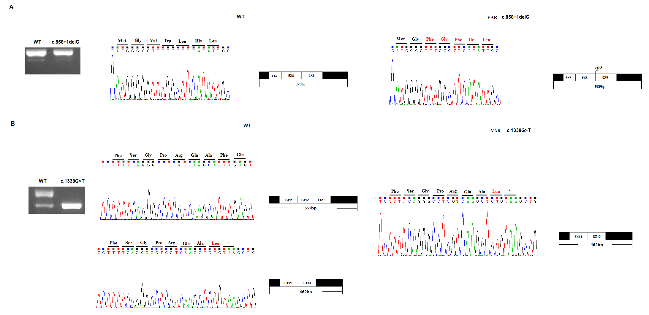
 Figure 3 of
Zhong, Mol Vis 2019; 25:204-214.
Figure 3 of
Zhong, Mol Vis 2019; 25:204-214.