Figure 5. Vascular endothelial growth factor (VEFG) and von Willebrand factor (vWF) immunofluorescence in retina/choroid sections from
PBS- and PEG-injected eyes at 14 days after injection. VEGF immunoreactivity (red) was evident in (A) a positive control sample from a human eyelid cavernous haemangioma, courtesy of the IOBA Ocular Pathology Lab, but was
not apparent in the retina/choroid of the (B) PBS- or (C) PEG-injected eyes. Similarly, vWF immunoreactivity (green) was evident in (D) a positive control sample from a human eyelid cavernous haemangioma, but was not apparent in the retina/choroid of the (E) PBS- or (F) PEG-injected eyes. Blue, DAPI-labelled nuclei; Ch, choroid; NR, neuroretina; PEG: polyethylene glycol. Scale bar: 25 µm.
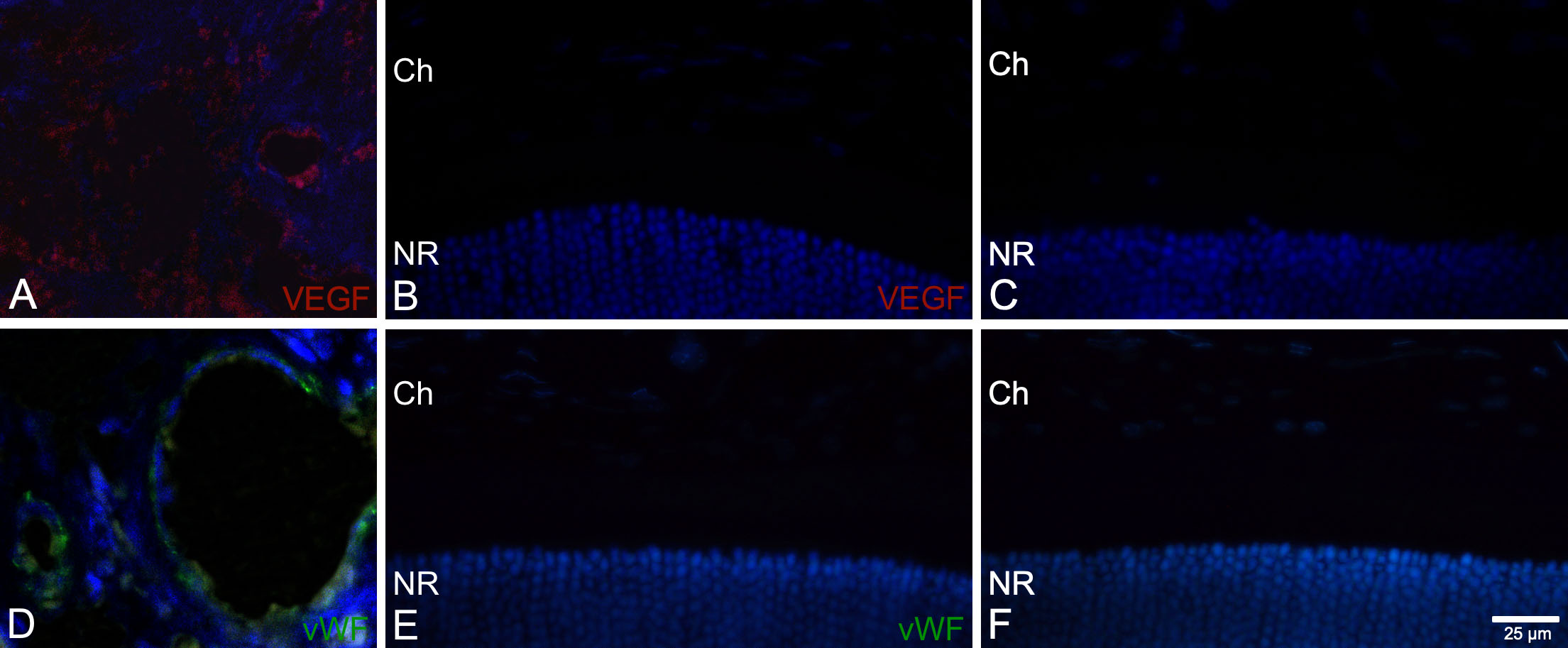
 Figure 5 of
Fernandez-Bueno, Mol Vis 2019; 25:194-203.
Figure 5 of
Fernandez-Bueno, Mol Vis 2019; 25:194-203.