Figure 4. Isolectin B4 (IB4) immunofluorescence of retina/choroid sections from PBS- and PEG-injected eyes at 14 days after injection.IB4
immunostaining (green) revealed morphological differences in the choroidal vessel pattern between (A) PBS- and (B) PEG-injected eyes (arrows). Vessel walls of the PEG-injected eyes appeared diffusely thickened. Blue, DAPI-labelled nuclei;
Ch, choroid; NR, neuroretina. Scale bar: 25 µm. C: Quantification of IB4 immunofluorescence in subretinal PBS- (white bars) and PEG-injected (black bars) mice. A.U.: arbitrary
units. *p<0.05; **p<0.001 between subretinal PBS- and PEG-injected eyes at different time points. PEG: polyethylene glycol.
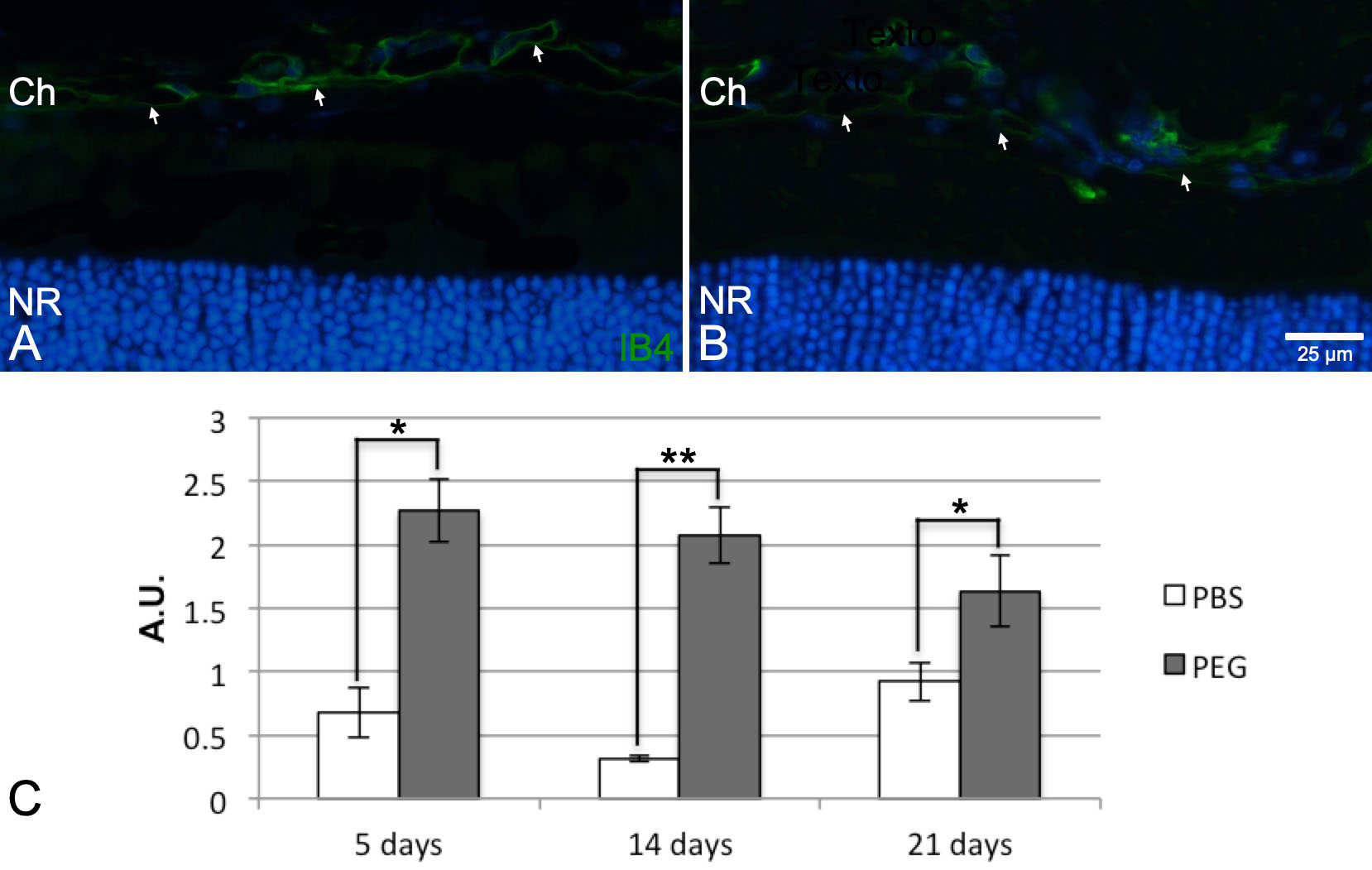
 Figure 4 of
Fernandez-Bueno, Mol Vis 2019; 25:194-203.
Figure 4 of
Fernandez-Bueno, Mol Vis 2019; 25:194-203.