Figure 3. Fontana-Masson (FM) staining and cytokeratin 8/18 (CK8/18) immunofluorescence in retina-choroid sections from mice at 14 days
after polyethylene glycol (PEG) injection. In these representative micrographs, (A) FM staining revealed the presence of melanin granules in the pigmented clumps at the subretinal space (arrowheads) and in
the RPE and choroid (arrows). Black: melanin; pink-red: nuclei and cytoplasm. B: In non-injected mouse eyes, immunostaining of CK8/18 (green) was present in the monolayer of RPE cells (arrows). C: In PEG-injected eyes, CK8/18 was present in cellular elements at the subretinal space (arrowheads), as well as in the RPE
cells (arrows). Asterisk: autofluorescence from photoreceptor outer segments; blue, DAPI-labelled nuclei. Ch: choroid; NR:
neuroretina. Scale bar: 25 µm.
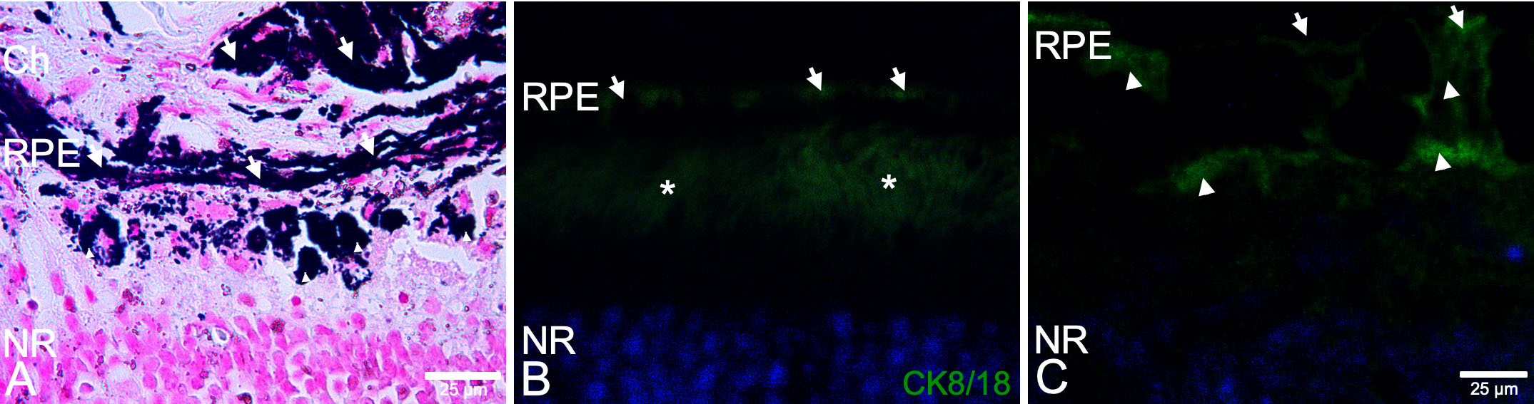
 Figure 3 of
Fernandez-Bueno, Mol Vis 2019; 25:194-203.
Figure 3 of
Fernandez-Bueno, Mol Vis 2019; 25:194-203.