Figure 2. Haematoxylin and eosin (HE) stained retina-choroid sections from PBS- and PEG (1 mg)-injected eyes. Representative micrographs
at 5 days after injection show (A) PBS-induced disruption of RPE/choroid cells (arrows) while (B) PEG-induced degeneration of photoreceptor outer segments (arrowhead), RPE cell enlargement, and vacuolization (asterisks).
At 14 days, (C) PBS-injected eyes presented subretinal vacuolization (asterisks) and disruption of the photoreceptor outer segments (arrowhead),
and (D) PEG-injected eyes revealed marked retinal degeneration and total RPE/choroid disruption (arrows). At 21 days, (E) PBS-injected eyes showed that the retina/choroid structures were preserved, while (F) PEG-injected eyes had RPE clumps and pigment cell migration (arrows). Ch: choroid; NR: neuroretina; PEG: polyethylene glycol.
Scale bar: 25 µm.
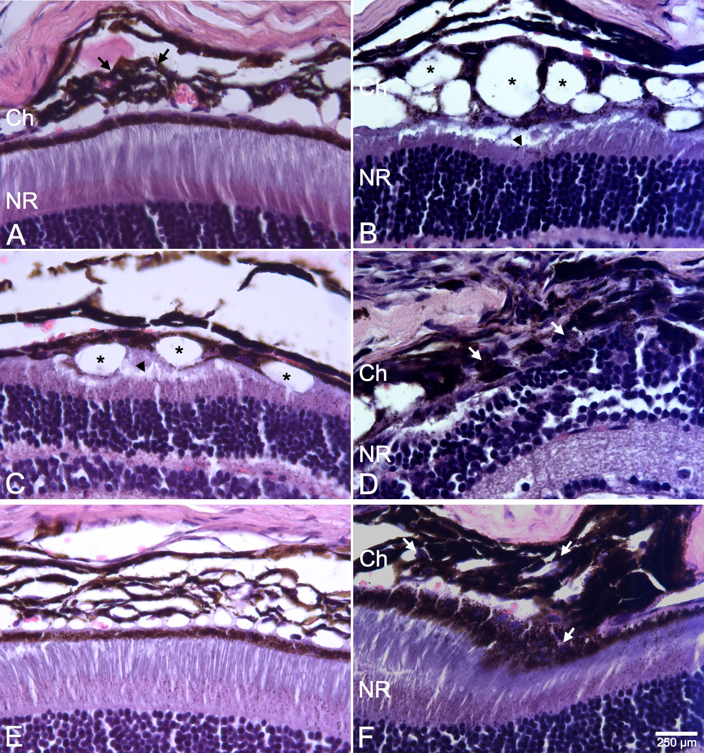
 Figure 2 of
Fernandez-Bueno, Mol Vis 2019; 25:194-203.
Figure 2 of
Fernandez-Bueno, Mol Vis 2019; 25:194-203.