Figure 1. Ophthalmic and histological appearance immediately after subretinal injections of PBS. A: Subretinal blebs visualized by surgical microscopy immediately after subretinal injection (arrows, delimitation of retinal
detachment extensions). B: Separation between the neuroretina and retinal pigment epithelium in the superior temporal quadrant of the eye (subretinal
injection area). No significant damage was evident in the neuroretina or in the retinal pigment epithelium/choroid. Photoreceptor
outer segment debris was present in the subretinal space (arrows). Haematoxylin and eosin staining, scale bar: 250 µm.
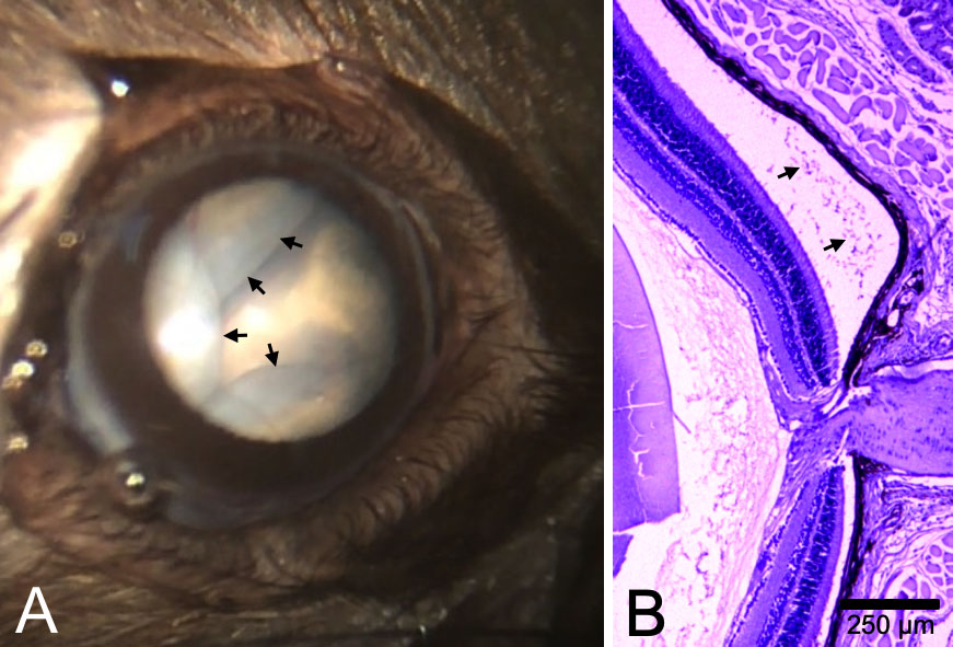
 Figure 1 of
Fernandez-Bueno, Mol Vis 2019; 25:194-203.
Figure 1 of
Fernandez-Bueno, Mol Vis 2019; 25:194-203.