Figure 2. Widefield autofluorescence and pseudocolor fundus imaging and SD–OCT of patient E-2. A, B: Autofluorescence and fundus Optos imaging at the age of 57 years, showing an atrophic macula, pigment deposits at the periphery,
and attenuated veins. C: Autofluorescence and optical coherence tomography (OCT) at the age of 54 years, showing central subfluid thickness on Heidelberg
spectral OCT due to severe edema causing elimination of the ellipsoid zone (EZ) temporally and at the foveal center in both
eyes. D: An inset picture from panel C, showing the magnified fovea and the lost EZ.
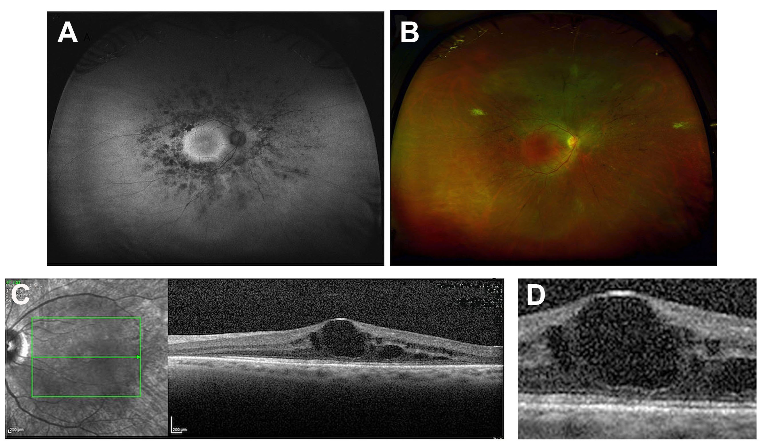
 Figure 2 of
Tatour, Mol Vis 2019; 25:155-164.
Figure 2 of
Tatour, Mol Vis 2019; 25:155-164.