Figure 6. Corneal findings using transmission electron microscopy of 2-month-old wild-type (WT; A and C) and Yap1+/− mice (B and D). A: The WT mice had a normal five- to eight-layer corneal epithelium, including discernable basal columnar cells with oval nuclei,
intermediate polygonal cells, and stratified squamous cells. B: The corneal epithelium in the Yap1+/− mice was thinner with two to four layers of cells, including a normal basal cell layer and a poorly differentiated superficial
squamous layer. The corneal stroma of the Yap1+/− mice was hypercellular with cells characterized by abundant cytoplasm, numerous variable-sized vacuoles, and oval nuclei,
interpreted as reactive keratocytes (black arrows). C: A single layer of flattened endothelial cells with well-structured gap junctions was identified resting on the normal Descemet’s
membrane in the WT mice. D: The Yap1+/− mice lacked a normal endothelium that was replaced by cuboidal melanocytes containing numerous pigment granules (melanin;
white arrowheads). These cells were observed overlying an absent or markedly thinned Descemet’s membrane. Epi, epithelium;
S, stroma; DM, Descemet’s membrane; E, endothelium.
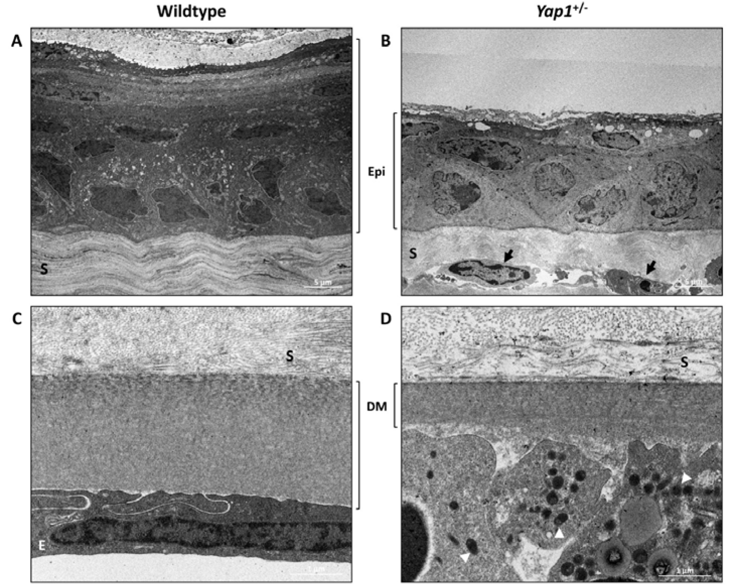
 Figure 6 of
Kim, Mol Vis 2019; 25:129-142.
Figure 6 of
Kim, Mol Vis 2019; 25:129-142.