Figure 5. Histopathology of cornea and retina in the Yap1+/- and WT mice with high magnifications. Normal corneal (A) and retinal (C) morphology was observed in 1-year-old wild-type (WT) mice. In the 1-year-old Yap1+/− mice, the cornea (B) was diffusely thinner than in the WT mice, presenting epithelial attenuation with loss of superficial squamous cells, and
hypercellular and compacted stroma with distorted lamellar arrangement and stromal neovascularization (*) and melanin pigmentation.
Descemet’s membrane was absent or severely thinned, and normal corneal endothelial cells were absent. In many locations, there
was fusion of the iris tissue to the exposed posterior cornea stroma, and melanotic cells lined the posterior cornea in many
regions where frank iris fusion was not observed (arrowheads). In 1-year-old Yap1+/− mice, retinal detachment was commonly observed and accompanied by subretinal accumulation of macrophages (arrows) and loss
of photoreceptors (D). Retinoschisis (*) of the inner nuclear layer and separation of the internal limiting membrane from the nerve fiber layer
(arrowheads) were observed. (A and B) 40X, hematoxylin and eosin (H&E); DM, Descemet’s membrane. (C and D) 20X, H&E; ILM, internal limiting membrane; GCL, ganglion cell layer; IPL, inner plexiform layer; INL, inner nuclear layer;
OPL, outer plexiform layer; ONL, outer nuclear layer; ELM, external limiting membrane; IS, inner segment; OS, outer segment;
RPE, retinal pigment epithelium.
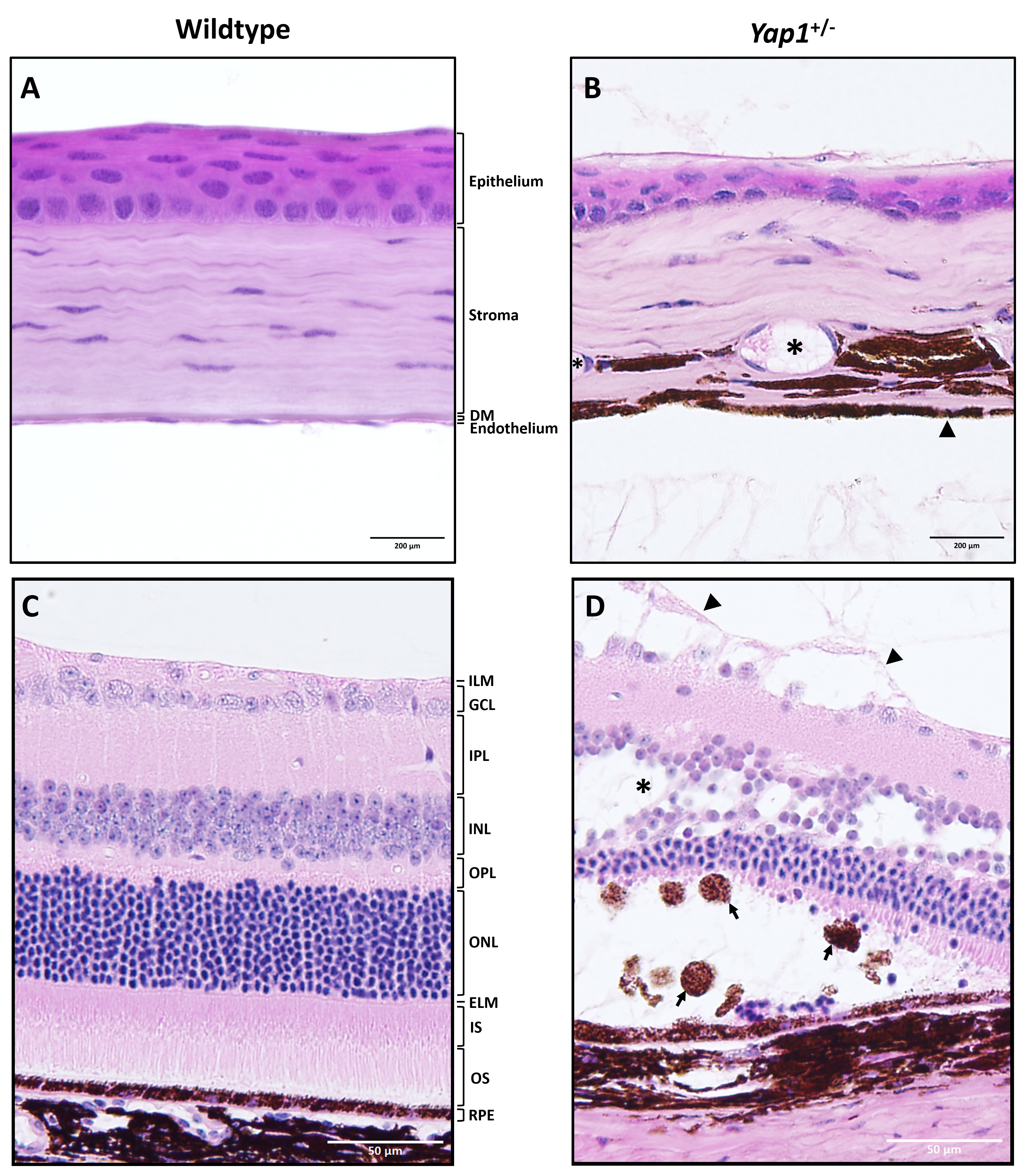
 Figure 5 of
Kim, Mol Vis 2019; 25:129-142.
Figure 5 of
Kim, Mol Vis 2019; 25:129-142.