Figure 3. Anterior segment images using optic coherence tomography (OCT) in Yap1+/- and WT mice. A: Fourier-domain optical coherence tomography demonstrates the normal anterior segment of male wild-type (WT) mice at the
indicated age: 2 weeks old, 2 months old, and 1 year old. B: The Yap1+/− mice demonstrate corneal pathology and anterior segment anomalies at all ages. A thin cornea with adherent iris (arrowhead),
a collapsed anterior chamber, and aphakia in age-matched male Yap1+/− mice at 2 weeks old, 2 months old, and 1 year old. A small lens and a deep anterior chamber were identified in the left eye
of a 1-year-old male Yap1+/− mouse. A, anterior chamber; C, cornea; I, iris; L, lens.
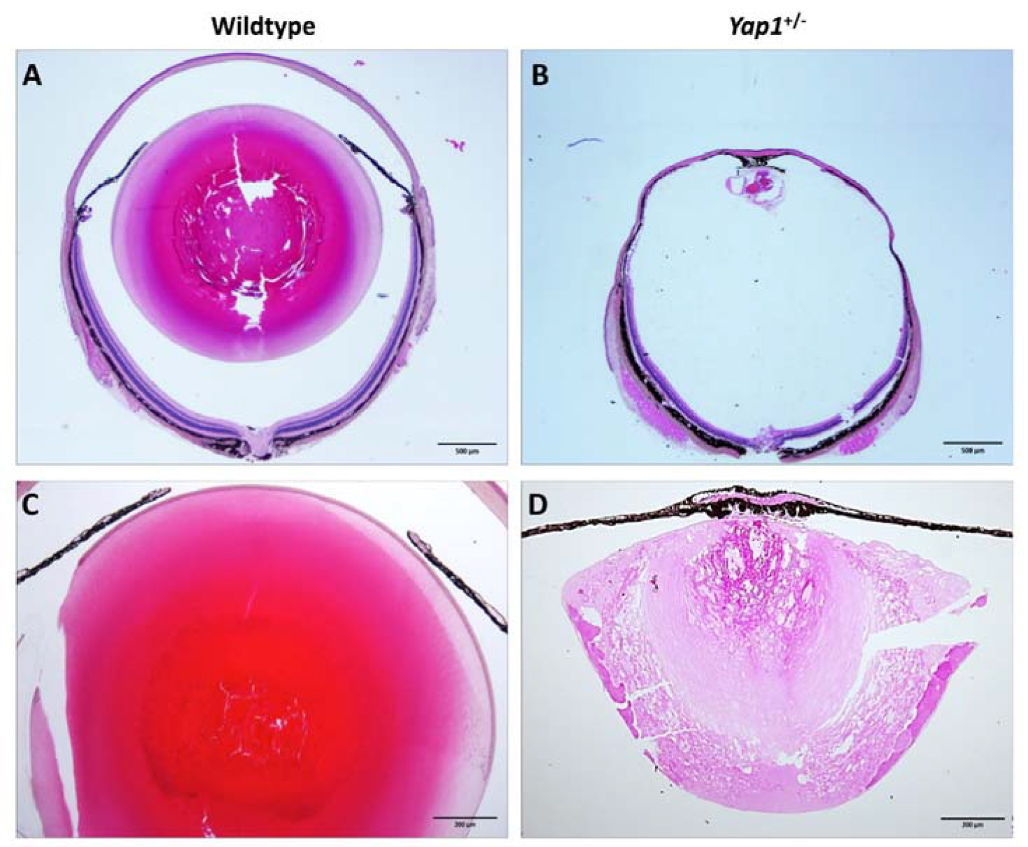
 Figure 3 of
Kim, Mol Vis 2019; 25:129-142.
Figure 3 of
Kim, Mol Vis 2019; 25:129-142.