Figure 2. YAP expression in Yap1+/- and WT mice. Immunofluorescent staining showed expression of YAP in the corneal epithelium and endothelium (A) and non-pigmented epithelium of the ciliary body (B) in wild-type (WT) and Yap1+/− mice. Decreased expression of YAP was identified in Yap1+/− versus WT mice.
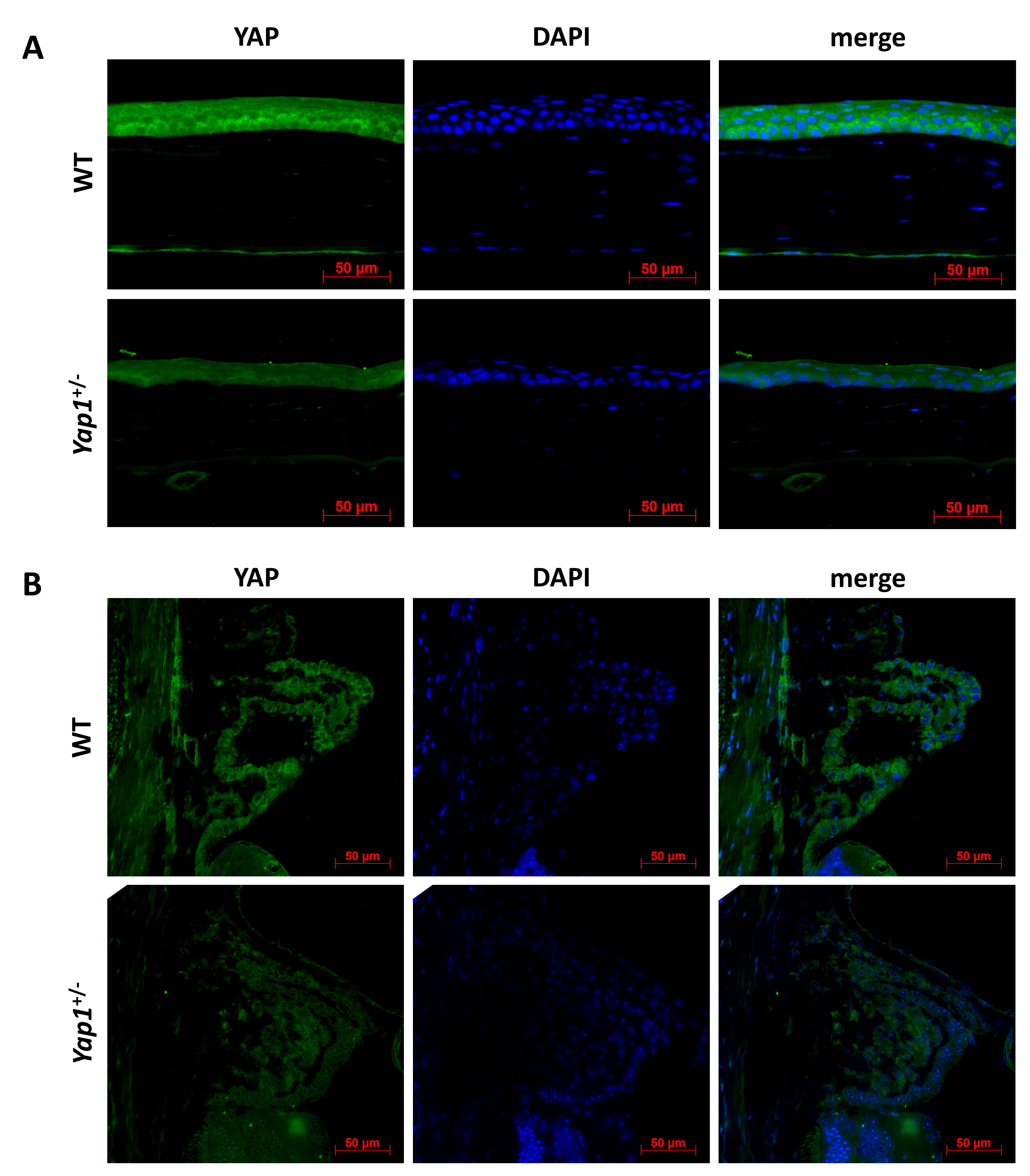
 Figure 2 of
Kim, Mol Vis 2019; 25:129-142.
Figure 2 of
Kim, Mol Vis 2019; 25:129-142.