Figure 1. Microphthalmia with anterior segment dysgenesis in Yap1+/- mice. Many Yap1+/− mice were microphthalmic with anterior segment dysgenesis in comparison to age-matched WT littermates. A: Compared with the wild-type (WT) mouse, microphthalmia with a small palpebral fissure was identified at all ages of Yap1+/− mice on gross examination. B: A normal transparent cornea with an appropriate anterior chamber, iris, and lens are found on biomicroscopic examination
with diffuse illumination (left) and narrow-slit beam (right) in the WT mice. Many Yap1+/− mice displayed generalized corneal fibrosis and a collapsed anterior chamber with the iris adherent to the posterior cornea
(left eye; 2 months old; right eye; 1 year old). A perforated cornea and granulation tissue were observed in the right eye
of a 1-year-old male Yap1+/− mouse. A hypermature cataract and posterior synechia with a deep anterior chamber were identified in the left eye of a 1-year-old
male Yap1+/− mouse with a transparent cornea.
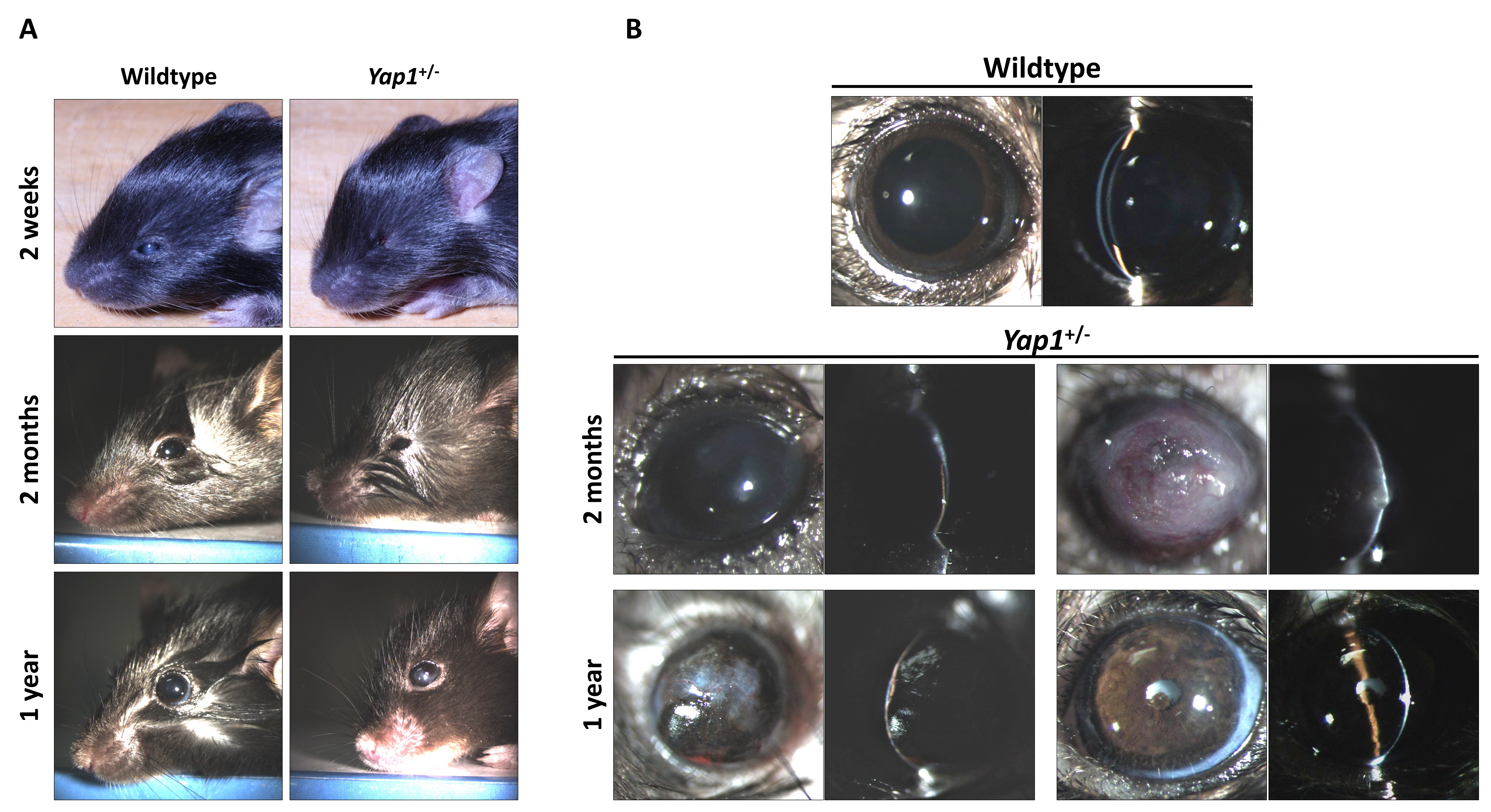
 Figure 1 of
Kim, Mol Vis 2019; 25:129-142.
Figure 1 of
Kim, Mol Vis 2019; 25:129-142.