Figure 5. 3D modeling of Gram-negative biofilms. Volumetric reconstruction with the surface function in Imaris software was used to
create a three-dimensional (3D) model of colonized bacteria. Green represents viable bacteria. Red represents non-viable bacteria
and extracellular DNA. A: Serratia marcescens, no neutrophils. B: S. marcescens with neutrophils. C: The S. marcescens biofilm volume was statistically significantly increased in the presence of neutrophils (p = 0.001, Student t test, n = 3). D: Pseudomonas aeruginosa, no neutrophils. E: P. aeruginosa with neutrophils. F: The biofilm volume of P. aeruginosa was statistically significantly increased in the presence of neutrophils compared to bacteria alone (p = 0.008, Student t test, n = 3). G: Stenotrophomonas maltophilia, no neutrophils. H: S. maltophilia with neutrophils. I: The biofilm volume of S. maltophilia was similarly increased in the presence of neutrophils (p<0.001, Student t test, n = 3). Scale bar = 20 µm.
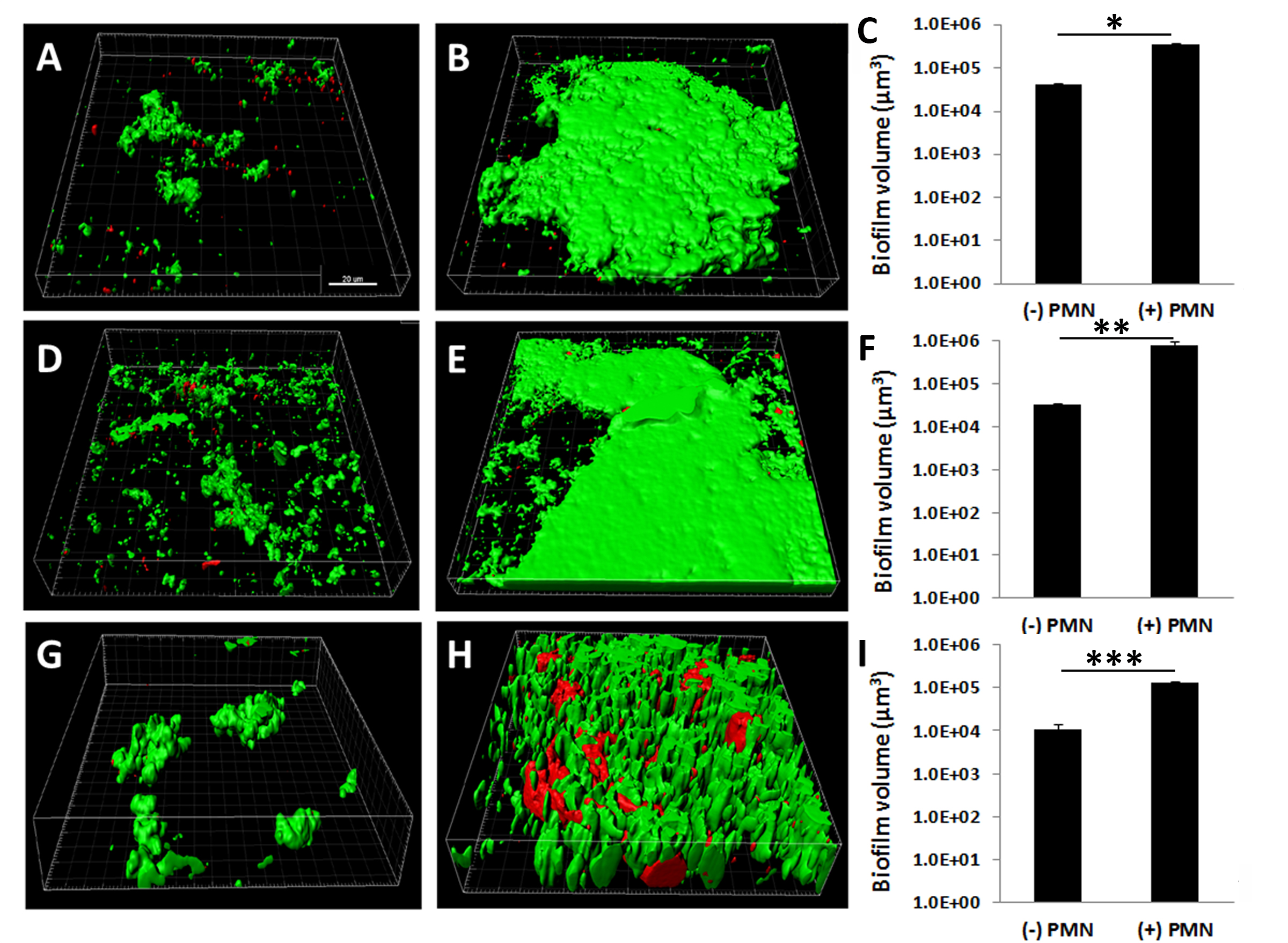
 Figure 5 of
Patel, Mol Vis 2018; 24:94-104.
Figure 5 of
Patel, Mol Vis 2018; 24:94-104.