Figure 4. 3D modeling of Gram-positive biofilms. Volumetric reconstruction with the surface function in Imaris software was used to
create a three-dimensional (3D) model of colonized bacteria. Green represents viable bacteria. Red represents non-viable bacteria
and extracellular DNA. A: Staphylococcus aureus, no neutrophils. B: S. aureus with neutrophils. C: The biofilm volume was statistically significantly increased in the presence of neutrophils (p = 0.010, Student t test, n = 3). D: Staphylococcus epidermidis, no neutrophils. E: S. epidermidis with neutrophils. F: There was no statistically significant difference in the biofilm volume between the neutrophils and the non-neutrophil control
(p = 0.0573, Student t test, n = 3). Scale bar = 20 µm.
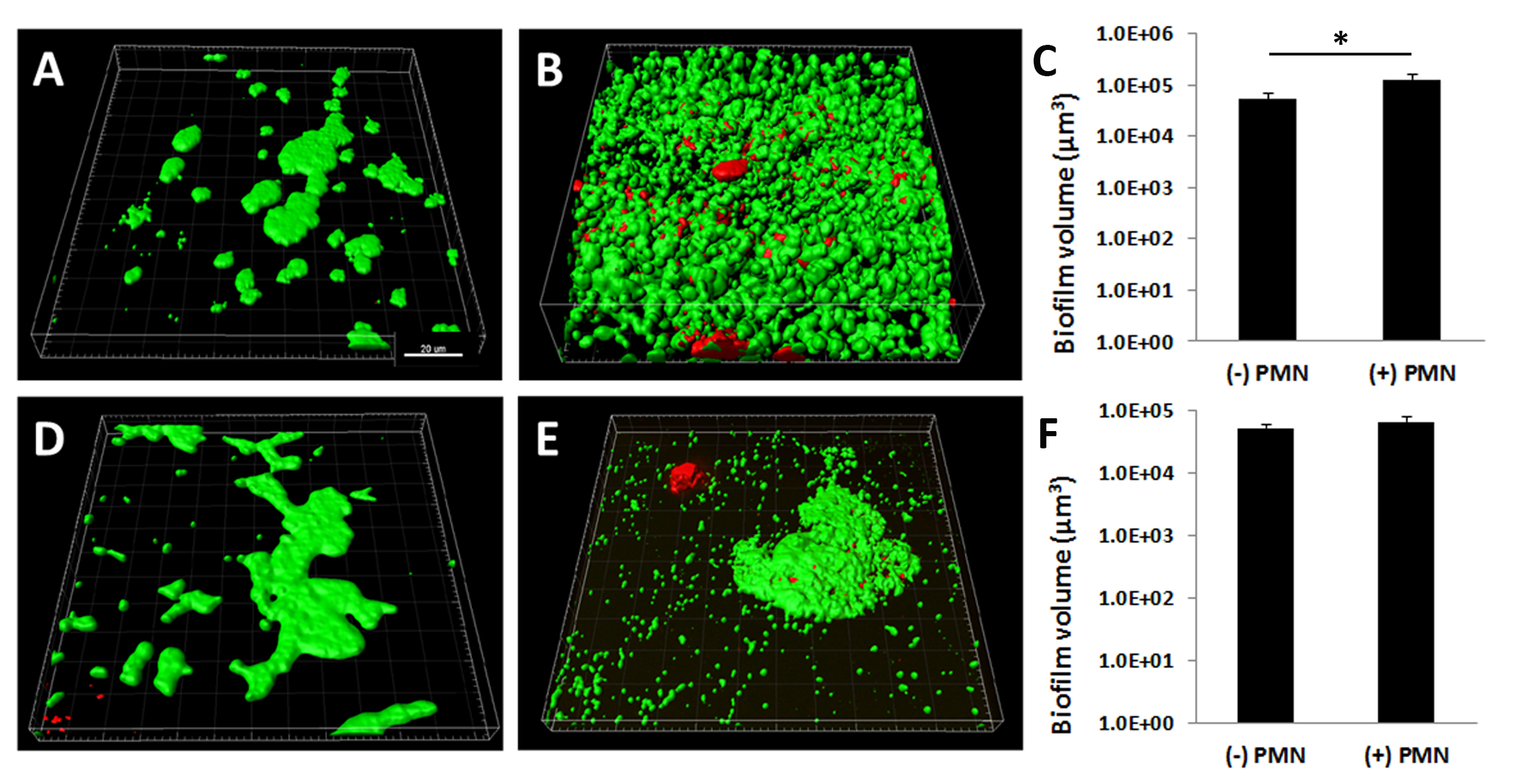
 Figure 4 of
Patel, Mol Vis 2018; 24:94-104.
Figure 4 of
Patel, Mol Vis 2018; 24:94-104.