Figure 3. Acceleration of Gram-negative bacterial colonization in the presence of neutrophils. A–D, I–J: Scanning electron microscopy of colonized bacteria. A: Serratia marcescens, no neutrophils. B: S. marcescens with neutrophils. C: Pseudomonas aeruginosa, no neutrophils. D: P. aeruginosa with neutrophils. I: Stenotrophomonas maltophilia, no neutrophils. J: S. maltophilia with neutrophils. Scale bar = 2 µm. E–H, K–L: BacLight staining of the bacteria adherent to the contact lens surfaces. Viable bacteria are shown in green and non-viable
bacteria and extracellular DNA in red. E: S. marcescens, no neutrophils. F: S. marcescens with neutrophils. G: P. aeruginosa, no neutrophils. H: P. aeruginosa with neutrophils. K: S. maltophilia, no neutrophils. L: S. maltophilia with neutrophils. Scale bar = 20 µm. M: Viable bacteria recovered from the contact lens surfaces. All three Gram-negative test strains showed a statistically significant
increase in viable bacteria adherent to the lens surface when incubated with neutrophils compared to the non-neutrophil control
(*p<0.001, two-way ANOVA, n = 3). Bacterial colonization was statistically significantly increased in the presence of neutrophils
with P. aeruginosa compared with the other two test strains (**p<0.001, two-way ANOVA, n = 3).
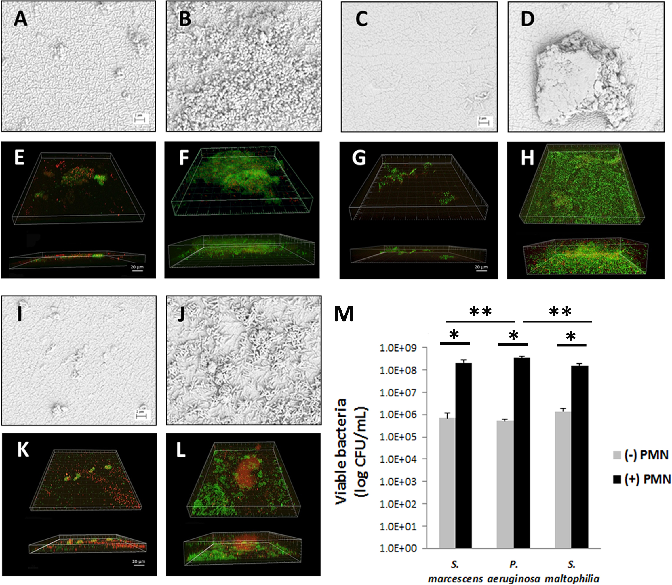
 Figure 3 of
Patel, Mol Vis 2018; 24:94-104.
Figure 3 of
Patel, Mol Vis 2018; 24:94-104.