Figure 2. Acceleration of Gram-positive bacterial colonization in the presence of neutrophils. A–D: Scanning electron microscopy of the colonized bacteria. A: Staphylococcus aureus, no neutrophils. B: S. aureus with neutrophils. C: Staphylococcus epidermidis, no neutrophils. D: S. epidermidis with neutrophils. Scale bar = 2 µm. E–F: BacLight staining of the bacteria adherent to the contact lens surfaces. Viable bacteria are shown in green and non-viable
bacteria and extracellular DNA in red. E: S. aureus, no neutrophils. F: S. aureus with neutrophils. G: S. epidermidis, no neutrophils. H: S. epidermidis with neutrophils. Scale bar = 20 µm. I: Viable bacteria recovered from the contact lens surfaces. S. aureus and S. epidermidis showed a statistically significant increase in viable bacteria adherent to the lens surface when coincubated with neutrophils
(*p<0.001 and **p = 0.008 for S. aureus and S. epidermidis, respectively; two-way ANOVA, n = 3). Neutrophil-mediated adhesion was greatest for S. aureus (*p<0.001, two-way ANOVA, n = 3).
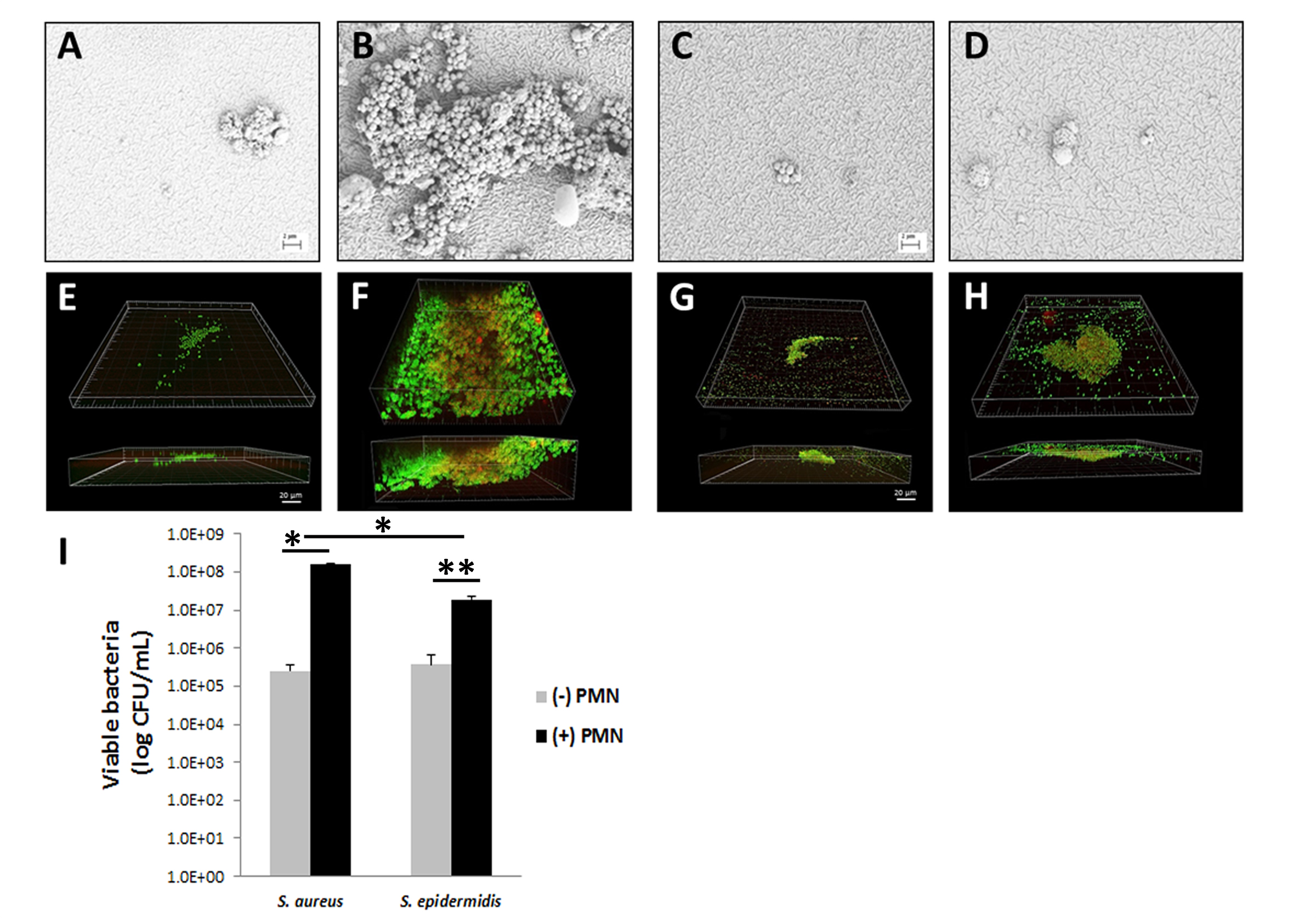
 Figure 2 of
Patel, Mol Vis 2018; 24:94-104.
Figure 2 of
Patel, Mol Vis 2018; 24:94-104.