Figure 5. Transmission electron microscopy micrograph of conjunctiva. A: A stromal telocyte between two vessels and Tps of the perivascular sheaths were observed. Homocellular contact points (A, B, arrows) and a TC/endothelial cell heterocellular junction (A, arrowhead and high magnification inset) were observed. Scale bar: A–B = 1 µm.
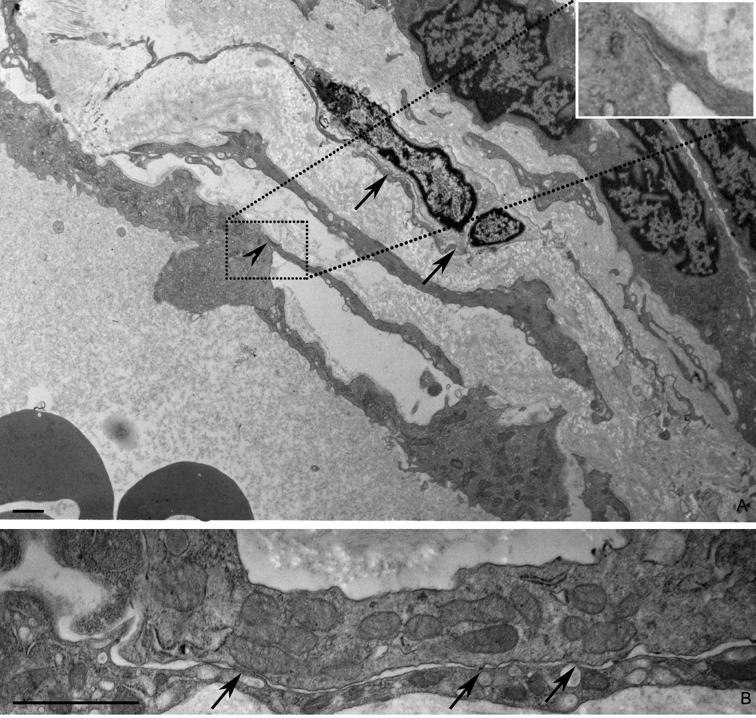
 Figure 5 of
Maxia, Mol Vis 2018; 24:853-866.
Figure 5 of
Maxia, Mol Vis 2018; 24:853-866.