Figure 3. Immunohistochemical analysis of TCs in pterygium and conjunctiva. A,B: C-kit. C: Vimentin. D: VEGF. E: S100. F: Laminin. G,H: α-SMA. I,J: CD133. K,L: CD31. A,C–G,I,K: pterygium. B,H,J,L: conjunctiva. In pterygium (A) and conjunctiva (B), telocytes were always negative for c-kit; scattered c-kit-immunoreactive cells, morphologically identifiable as mast cells,
were observable in the lamina propria. In pterygium, stromal TCs showed moderate immunostaining to vimentin (C, arrows) and VEGF (D, arrows); weak staining to S100 (E, arrows) and laminin (F, arrows) in a few stromal TCs was observed. G,H: Only adventitial TCs of the perivascular sheaths showed evident staining to α-SMA. I: CD133 immunoreactivity in pterygium TCs was completely absent and limited to the endothelial cells of neoformed blood vessels.
No immunostaining in conjunctiva was observed (J). In pterygium (K) and conjunctiva (L), only endothelial cells showed immunoreactivity to CD31. Scale bar: A–L = 50 µm.
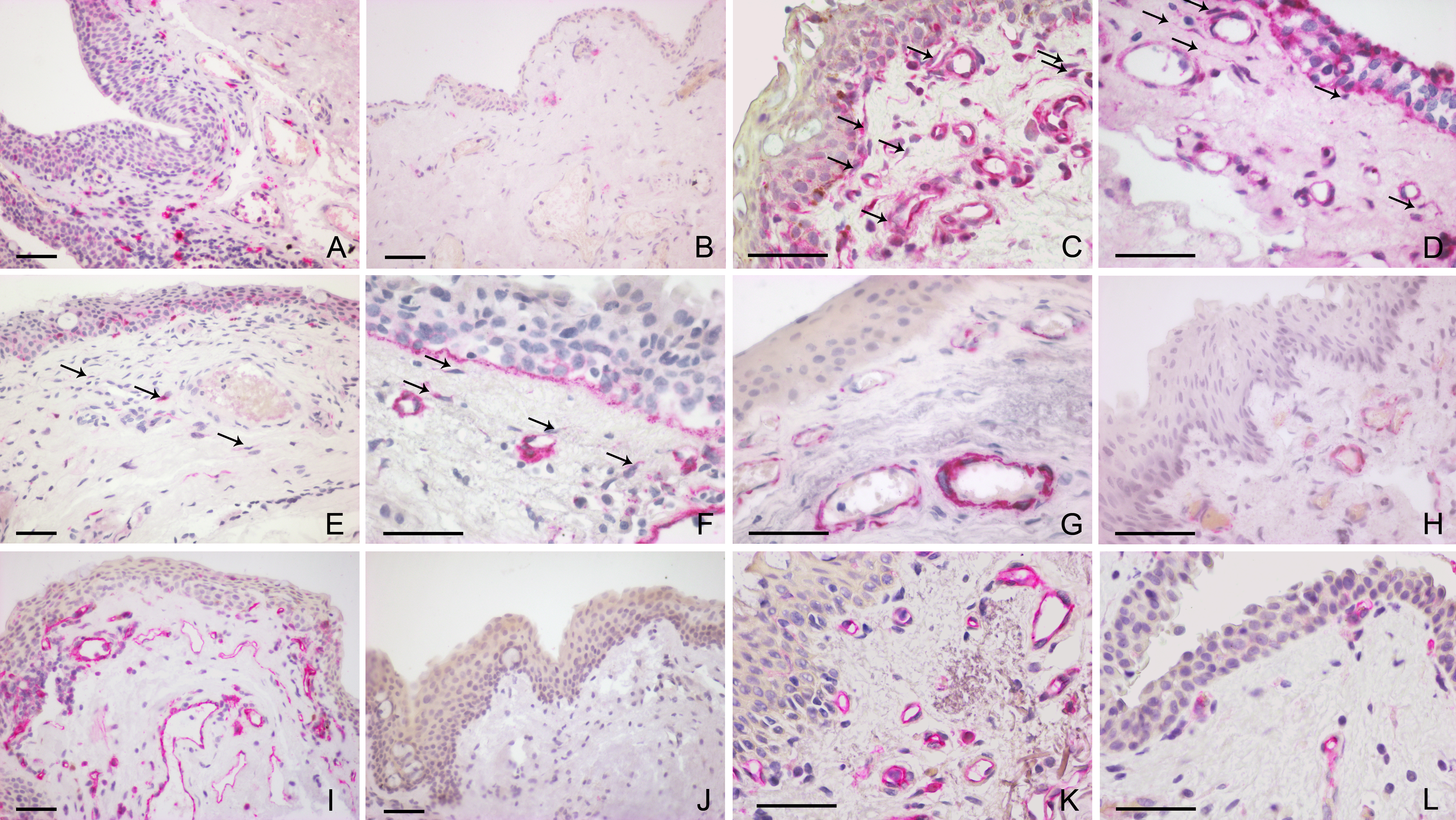
 Figure 3 of
Maxia, Mol Vis 2018; 24:853-866.
Figure 3 of
Maxia, Mol Vis 2018; 24:853-866.