Figure 2. Double immunofluorescence labeling of TCs in pterygium and conjunctiva. A-D: double immunofluorescent reaction for PDGFRα (green) and nestin (red); E-H: double fluorescence immunolabeling for PDGFRα (green) and CD34 (red). Extensive colocalization (yellow) of PDGFRα and nestin
(C) and PDGFRα and CD34 (G) in pterygium stromal telocytes was observed. Nestin was always expressed in a punctate/granular pattern (B,C). The scattered conjunctival stromal TCs always showed a PDGFRα+/nestin- (D) and PDGFRα+/CD34+ (H, arrows) immunophenotype. Nuclei (blue) are counterstained with 4′,6-diamidino-2-phenylindole (DAPI). Scale bar: A–H = 125 µm.
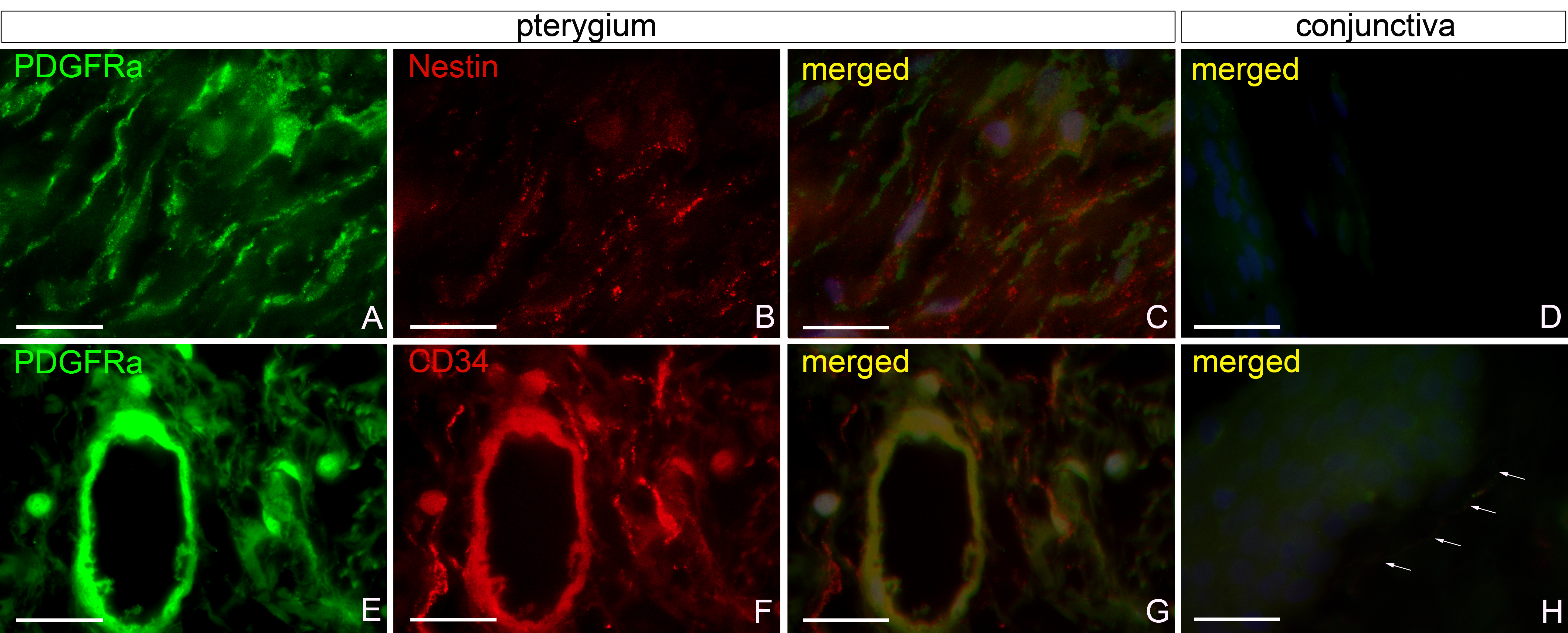
 Figure 2 of
Maxia, Mol Vis 2018; 24:853-866.
Figure 2 of
Maxia, Mol Vis 2018; 24:853-866.