Figure 1. Distribution of PDGFRα-, CD34-, and nestin-immunoreactive TCs in pterygium and conjunctiva. A–D: anti-human PDGFRα. E–H: anti-human CD34. I–L: anti-human nestin. Pterygium and conjunctiva shared the same telocyte distribution pattern; however, in pterygium marked
stromal TC hyperplasia was observed (A,E,J). In pterygium, strong immunoreactivity to PDGFRα, CD34, and nestin was localized underneath the epithelium along the basement
membrane (A,E,F,I,K). In the stroma, immunoreactive TCs were widespread and arranged in perivascular sheaths around the newly formed vessels
(A,B,E–G,J), as well as scattered in the stroma, where the TCs ran in bundles, parallel to the epithelium and simulating the course
of elastic fibers (A,E,J). C: In the fibrotic areas, TCs were almost absent, but on the border of the cicatricial area, stellate PDGFRα immunoreactive
TCs, with at least five extremely long, branched, dendritic-like processes, were detectable (arrows). E: The organization in parallel bundles went missing and was replaced with a wide TC network (arrows). K: High magnification of a nestin-positive TC, with very long telopodes, running along the basement membrane, was detectable.
In conjunctiva, mild immunoreactivity to PDGFRα (D) and CD34 (H) was observed. No immunostaining to nestin (L) was detected in any cases. Scale bar: A–J,L = 50 µm; K = 20 µm.
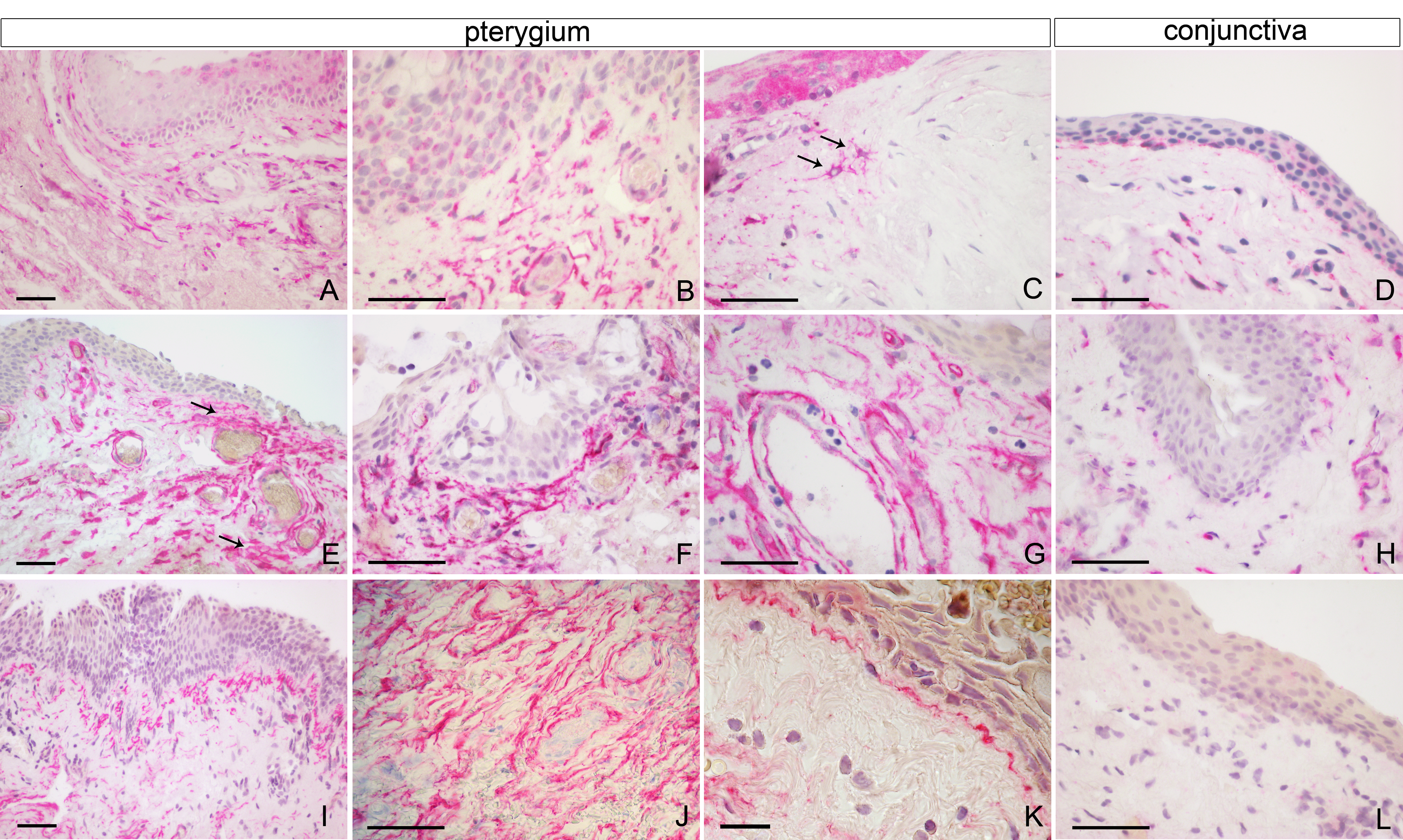
 Figure 1 of
Maxia, Mol Vis 2018; 24:853-866.
Figure 1 of
Maxia, Mol Vis 2018; 24:853-866.