Figure 6. COS proteins restored to the outer segments of treated Nphp5-/-; Nrl-/- mice. A–J: Immunohistochemistry of retina cryosections from untreated (left) and treated (right) Nphp5+/−; Nrl-/- (A, C, E, G, and I) and Nphp5-/-; Nrl-/- (B, D, F, H, and J) mice. Cryosections were probed with antibodies directed against S-opsin (A, B), ML-opsin (C, D), cone PDE6 (E, F), cone transducin-γ (G, H), and cone transducin-α (I, J). Lower panels (A–J) show enlargements of dashed boxes (yellow); all sections were from mice expressing Egfp-Cetn2+ (eCetn2+, green). Scale bar, 20 µm. Note the absence of cone phototransduction components in the untreated double-knockout (B, D, F, H, and J, left panels) and partial restoration following treatment (B, D, F, H, and J, right panels).
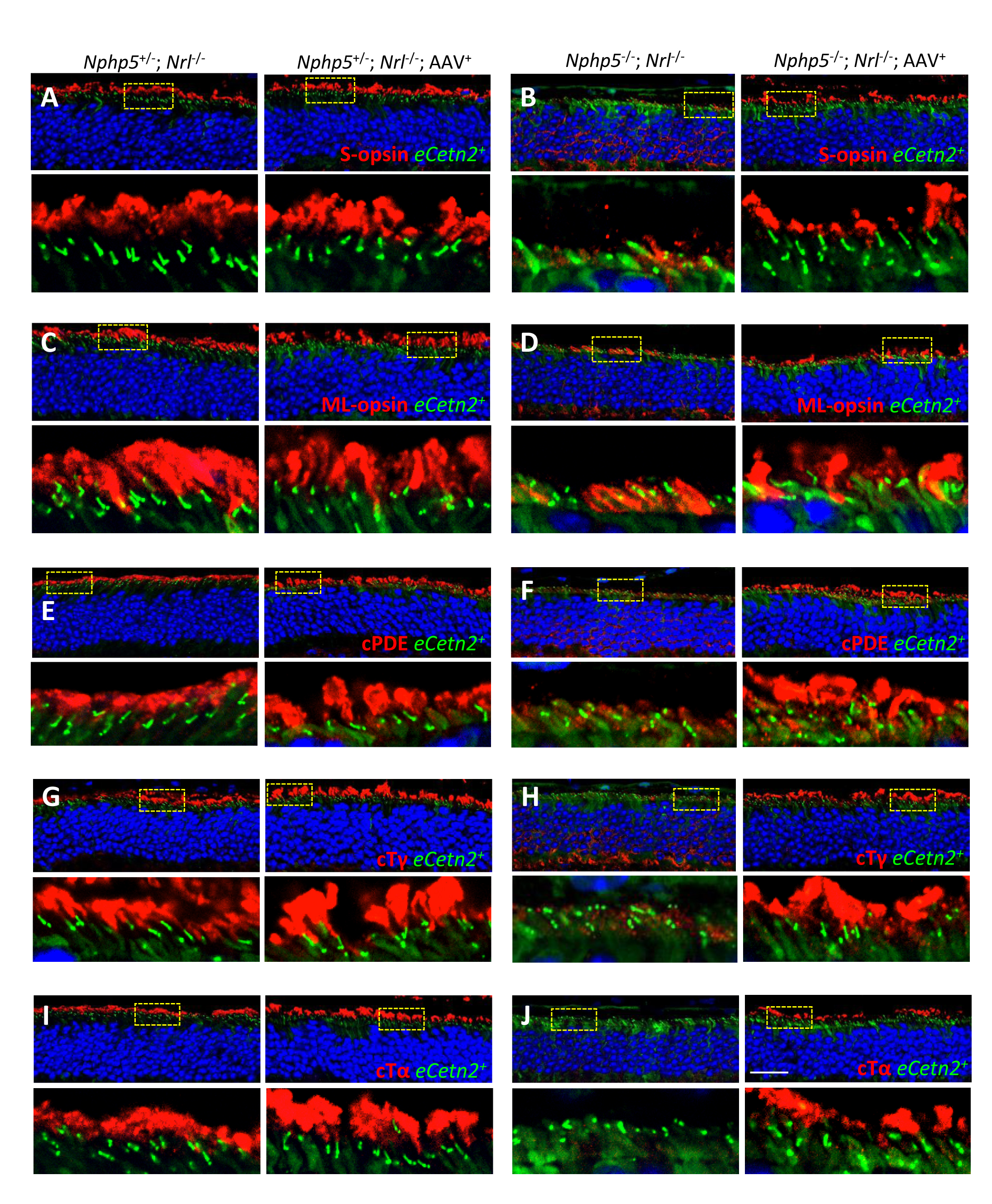
 Figure 6 of
Hanke-Gogokhia, Mol Vis 2018; 24:834-846.
Figure 6 of
Hanke-Gogokhia, Mol Vis 2018; 24:834-846.