Figure 5. scAAV8-FL-cNPHP5 treatment (AAV+) reinitiates cone ciliogenesis in Nphp5−/−; Nrl−/− retina. A, B: Immunohistochemistry of untreated (left) and treated (adenoassociated virus (AAV+), right) retina cryosections from Nphp5+/− (A), and Nphp5-/- mice (B). C, D: Immunohistochemistry of untreated (left) and treated (right) retina cryosections from Nphp5+/−; Nrl-/- (C) and Nphp5-/-; Nrl-/- (D) mice. Mutant mice were kept on the Egfp-Cetn2+ background (eCetn2+, green), and sections were probed with anti-RP1 antibody (red) to label the developing axoneme. Lower panels are enlargements
of hatched boxes. Note the presence of the axoneme (red), connecting cilium (CC, green, white arrows), and mother and daughter
centrioles (green dots) in the controls (C); the absence of axoneme and CC in the double-knockout mice (D, left panel); and reemergence of the axoneme, CC, and centrioles in the treated double-knockout mice (D, right panel). Cryosections in A–J are from mice on the Egfp-Cetn2+ background. Scale bar, 20 µm.
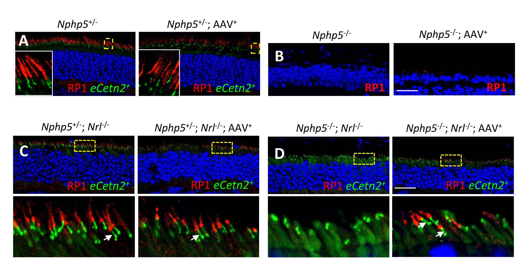
 Figure 5 of
Hanke-Gogokhia, Mol Vis 2018; 24:834-846.
Figure 5 of
Hanke-Gogokhia, Mol Vis 2018; 24:834-846.