Figure 4. Rescue of ciliogenesis with scAAV8 vector expressing cNPHP5, detected with immunohistochemistry using anti-Flag antibody.
A–D: Retina cryosections from Nphp5+/−; Nrl-/- (A), Nphp5+/−; Nrl-/- treated with self-complementary adenoassociated virus 8 (scAAV8)-cNPHP5 (AAV+; B), Nphp5-/-; Nrl-/- (C), and Nphp5−/−; Nrl−/−; AAV+ (D) were probed with monoclonal anti-Flag antibody (red). Lower panels show enlargements of hatched boxes. All mice expressed
the Egfp-Cetn2+ transgene (eCetn2+, green) which specifically labels centrioles and transition zones. Please note that Egfp-Cetn2+ binding to the lumen of NPHP5−/−; Nrl−/− centrioles is impaired resulting in dispersion to the inner segments. OS, outer segment; CC, connecting cilium; ONL, outer
nuclear layer. Scale bar, 20 µm. E–H: Cone PDE6 expression profile in untreated and treated (AAV+) retinas. Nphp5+/−; Nrl−/− (E), Nphp5−/−; Nrl−/− (F), Nphp5+/−; Nrl−/−; AAV+ (G), and Nphp5−/−; Nrl-/; AAV+ retina whole cryosections (H) were probed with anticone PDE6 (cPDE) antibody (red). Note the absence of expression in the double-knockout (F) and partial expression of cone PDE6 nasally in the treated retina. Indentation *, point of injection. Cryosections in A, C, and D are from mice on the Egfp-Cetn2+ background. Scale bar, 200 µm; enlargement, 50 µm.
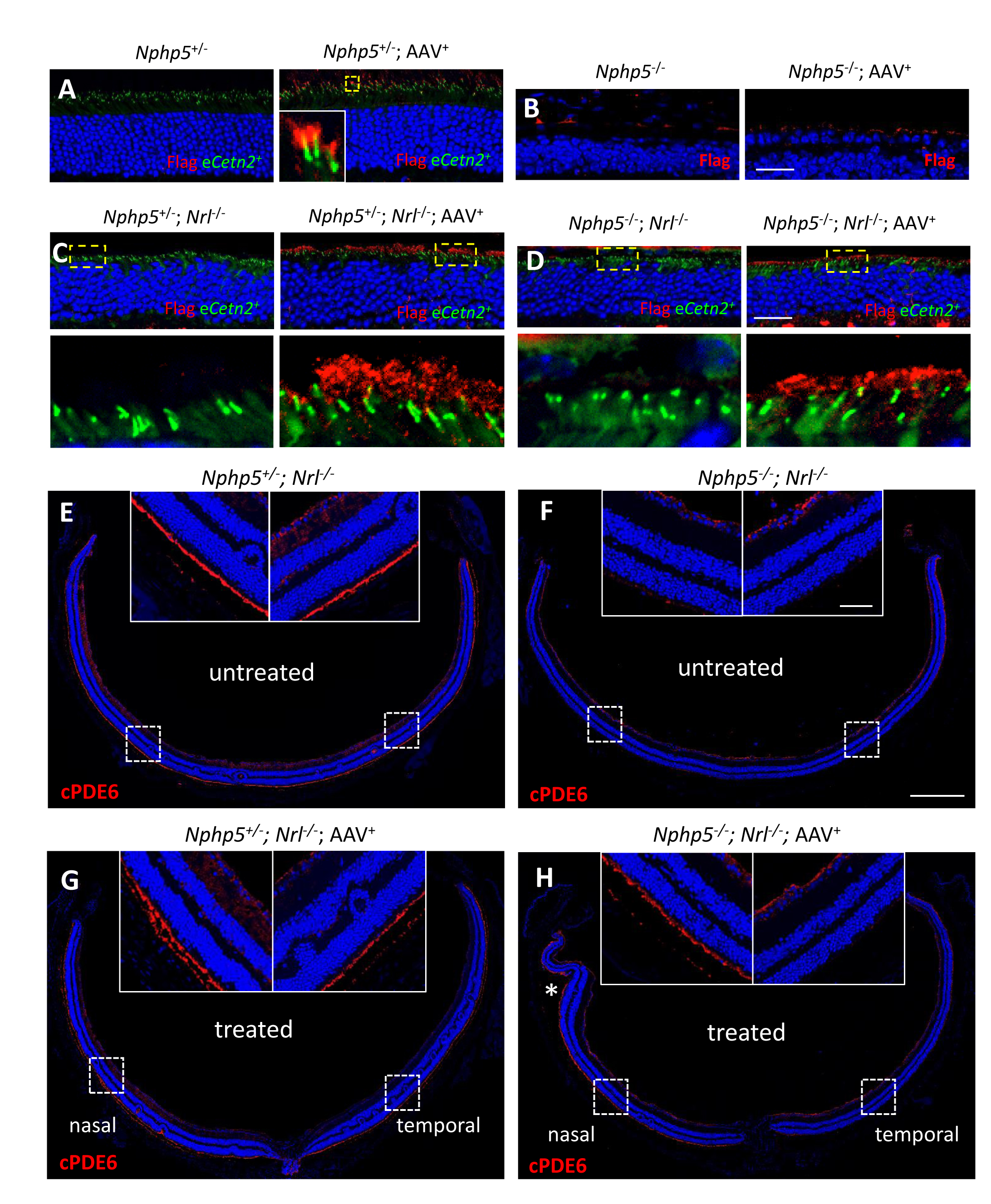
 Figure 4 of
Hanke-Gogokhia, Mol Vis 2018; 24:834-846.
Figure 4 of
Hanke-Gogokhia, Mol Vis 2018; 24:834-846.