Figure 4. YAP siRNA inhibits the proliferation of endothelial cells. A: The PCNA protein level was detected with western blotting. GAPDH was used as a loading control. B: The Bar Chart showed the ratio of PCNA to GAPDH. *p<0.05, compared to the normal control group. C: The PCNA protein level following scramble siRNA or YAP siRNA intravitreal (IP) injection was detected with western blotting.
GAPDH was used as a loading control. D: The Bar Chart showed the ratio of PCNA to GAPDH. *p<0.05, compared to the 7 day post-laser photocoagulation group. E: Double immunostaining of PCNA and CD31 in the normal control, 7 day post-laser photocoagulation, and YAP siRNA groups. Scale
bar = 100 μm.
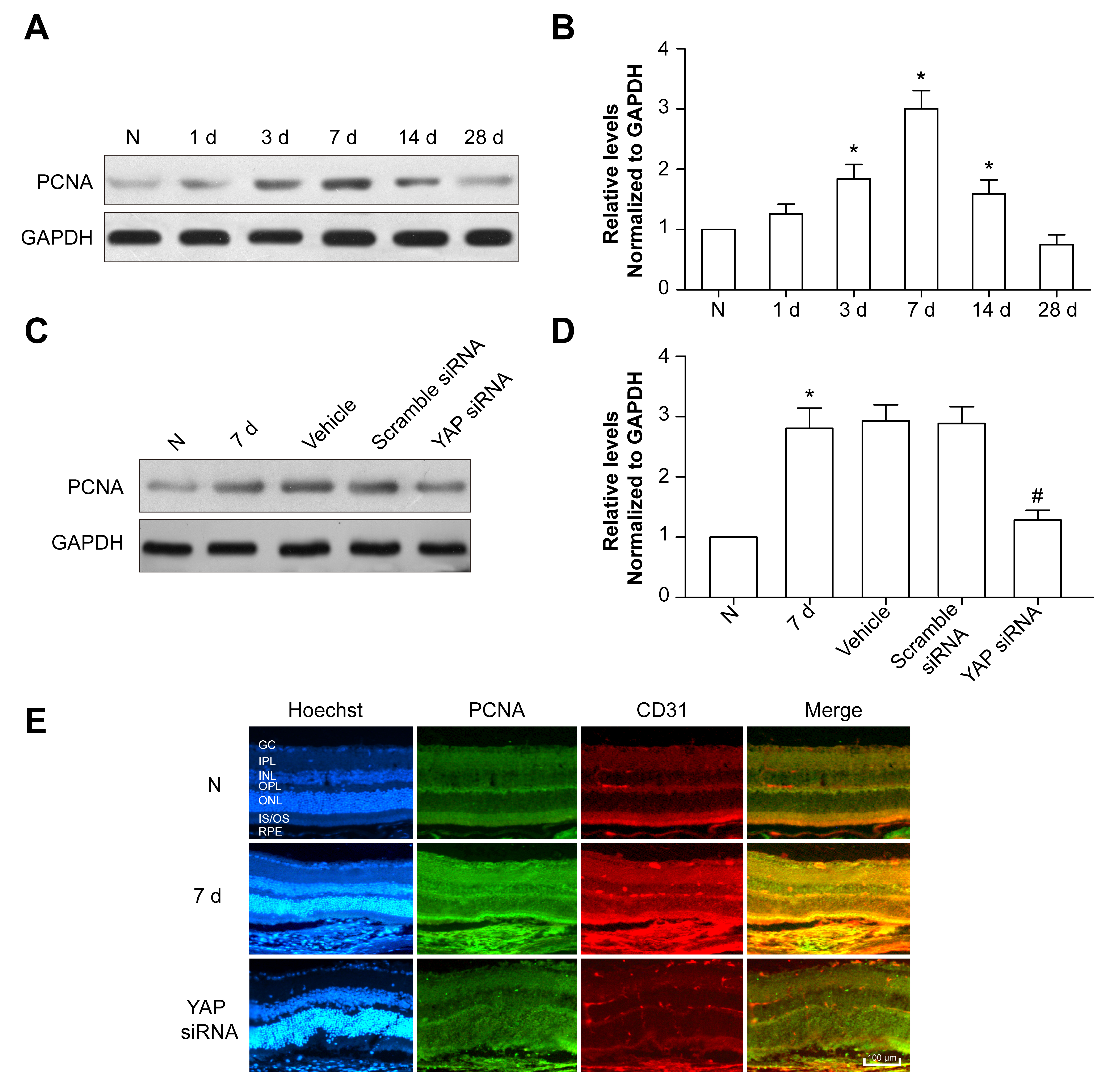
 Figure 4 of
Yan, Mol Vis 2018; 24:83-93.
Figure 4 of
Yan, Mol Vis 2018; 24:83-93.