Figure 3. YAP is localized in endothelial cells. A: Normal mouse retinal and choroid structure. Each photograph shows the central area of choroidal neovascularization (CNV)
within the mouse retinas and choroids in the 7 day post-laser photocoagulation, vehicle, scramble siRNA, YAP siRNA, or ranibizumab
(RBZ) group. Scale bar: 100 μm (RPE: retinal pigment epithelium; OS: outer segment; IS: inner segment; ONL: outer nuclear
layer; OPL: outer plexiform layer; INL: inner nuclear layer; IPL: inner plexiform layer; GC: ganglion cell layer). B: Statistical analysis of the data of the 7 day post-laser photocoagulation, vehicle, scramble siRNA, YAP siRNA, and RBZ groups.
n = 12–16 spots. *p<0.05, compared to the 7 day post-laser photocoagulation group. Black dashed lines represent the edge of
CNV. C: Cellular localization of YAP in the retina/choroid cryosections was determined with double immunostaining with endothelial
marker CD31. Scale bar = 100 μm.
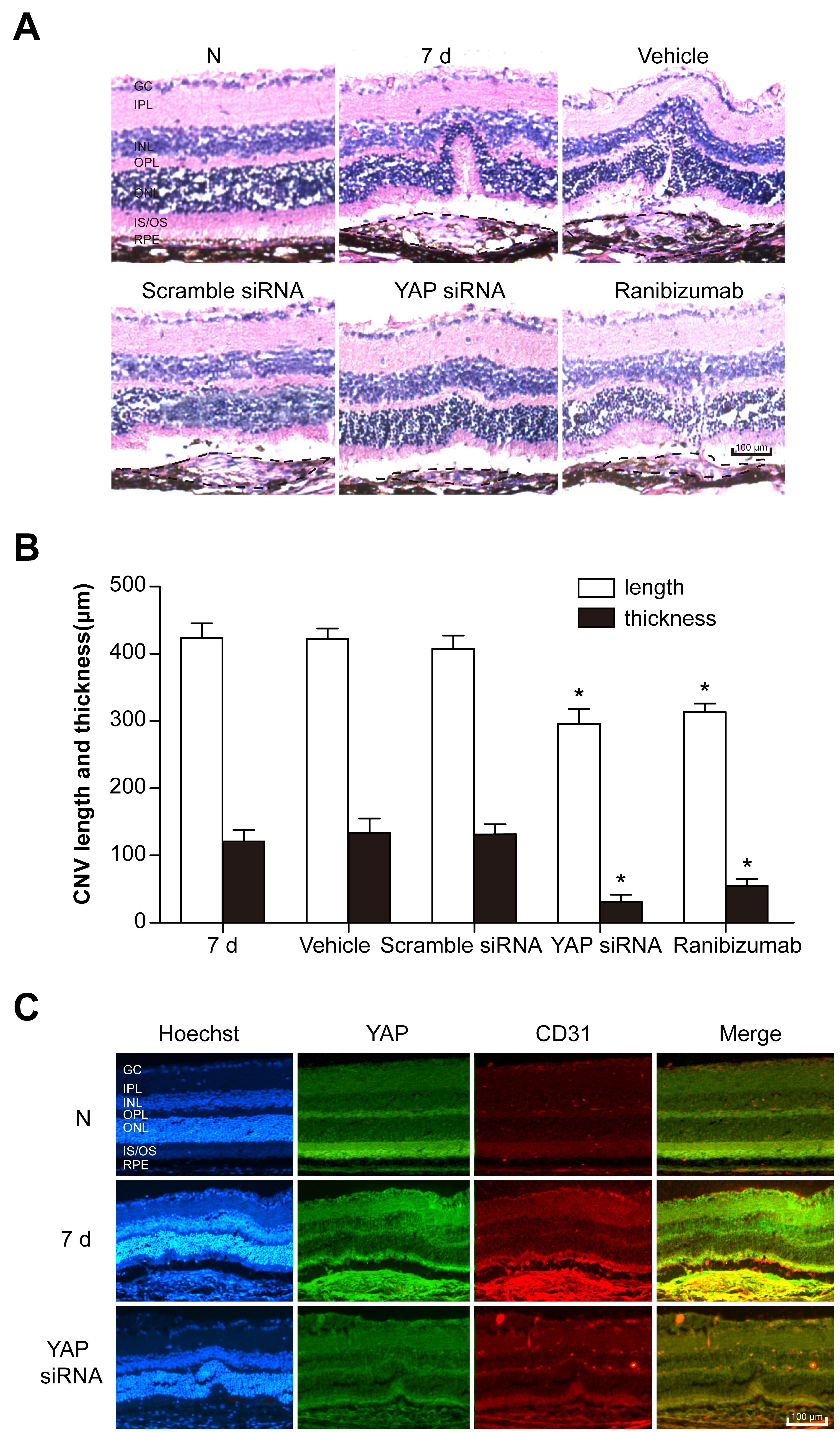
 Figure 3 of
Yan, Mol Vis 2018; 24:83-93.
Figure 3 of
Yan, Mol Vis 2018; 24:83-93.