Figure 1. YAP and VEGF expression levels are upregulated after CNV formation. The mouse choroidal neovascularization (CNV) model was
generated by laser photocoagulation. A: YAP and VEGF protein levels were detected with western blotting. GAPDH was used as a loading control. B: The histogram shows the densitometric analysis of the average levels of YAP and VEGF relative to GAPDH. *p<0.05; compared
to normal controls. C: Immunohistochemistry of YAP in the normal and 7 day post-laser photocoagulation groups.
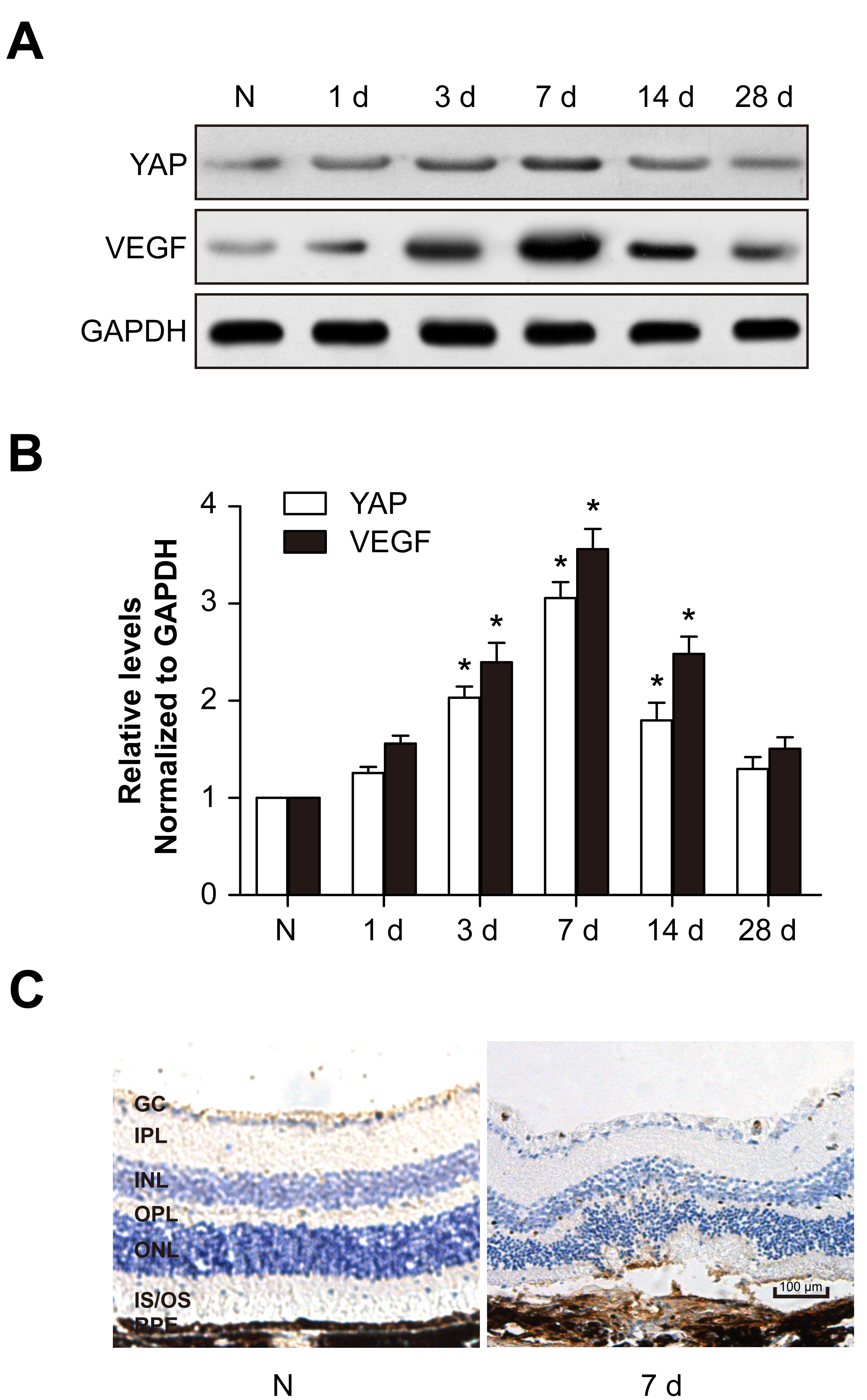
 Figure 1 of
Yan, Mol Vis 2018; 24:83-93.
Figure 1 of
Yan, Mol Vis 2018; 24:83-93.