Figure 4. WAXS polar vector map showing preferential collagen orientation across non-myopic flat-mounted posterior sclera N4, overlaid
over a photograph of the tissue before scanning. The superior direction of the specimen is indicated with a blue arrow. Polar
vectors are color coded according to the bar, with warmer colors indicating higher degrees of collagen anisotropy. Note the
highly aligned collagen annulus circumscribing the nerve head (the black line bounded region), two tangential fiber bands
(black arrows), and the uniaxial alignment of the ocular muscle insertion regions, with the inferior oblique highlighted (red
arrow).
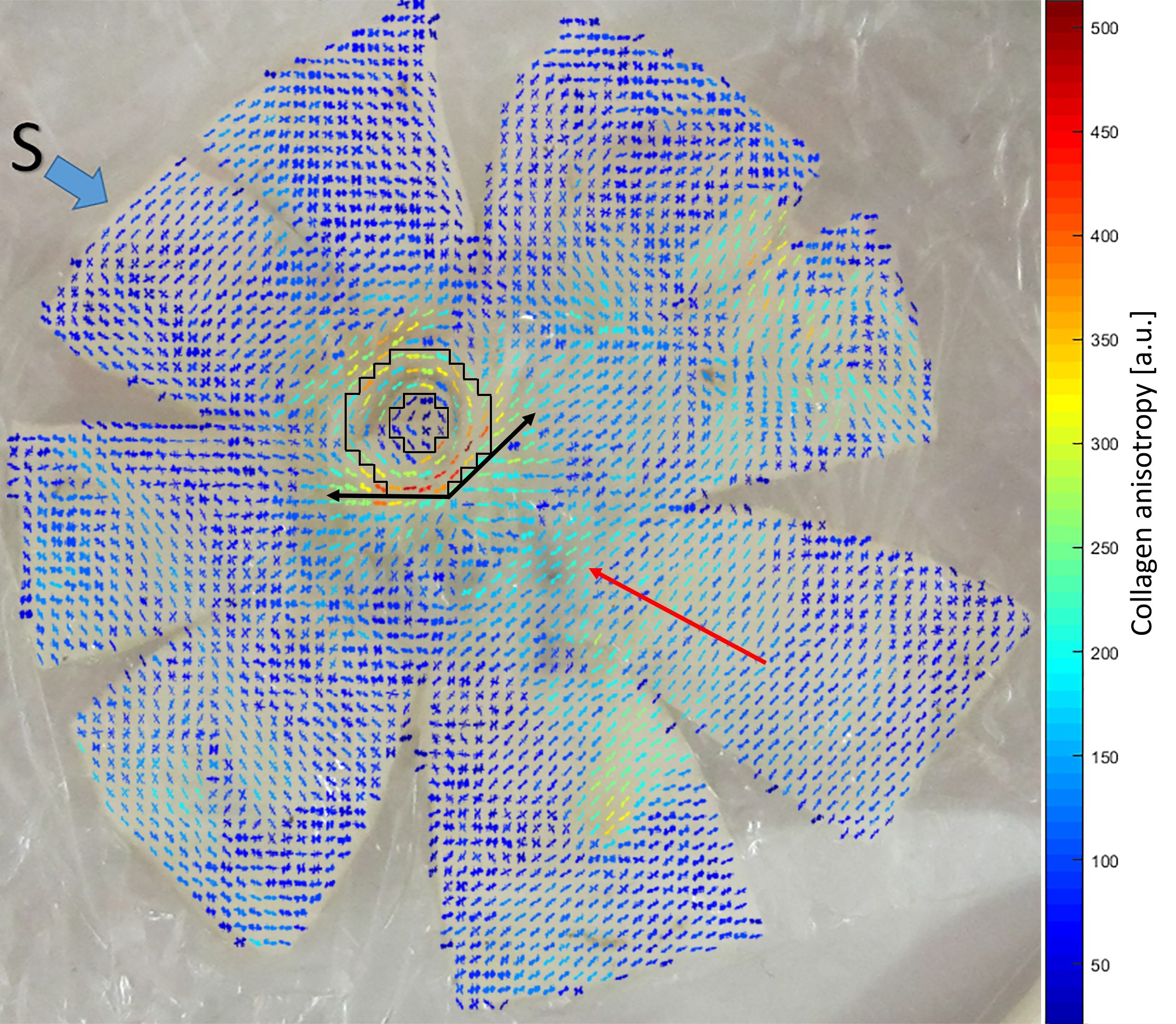
 Figure 4 of
Markov, Mol Vis 2018; 24:818-833.
Figure 4 of
Markov, Mol Vis 2018; 24:818-833.