Figure 2. Beamline I03 at the Diamond Light Source operating in a custom fiber-diffraction set-up. The goniometer (A) provides directional translation of the sample holder (B) between X-ray exposures. A flat-mounted posterior sclera is shown mounted between Mylar sheets. After the specimen is positioned,
an additional Mylar sheet (C) in which a lead beam stop (D) is attached prevents undiffracted X-rays from reaching and damaging the detector positioned out of shot.
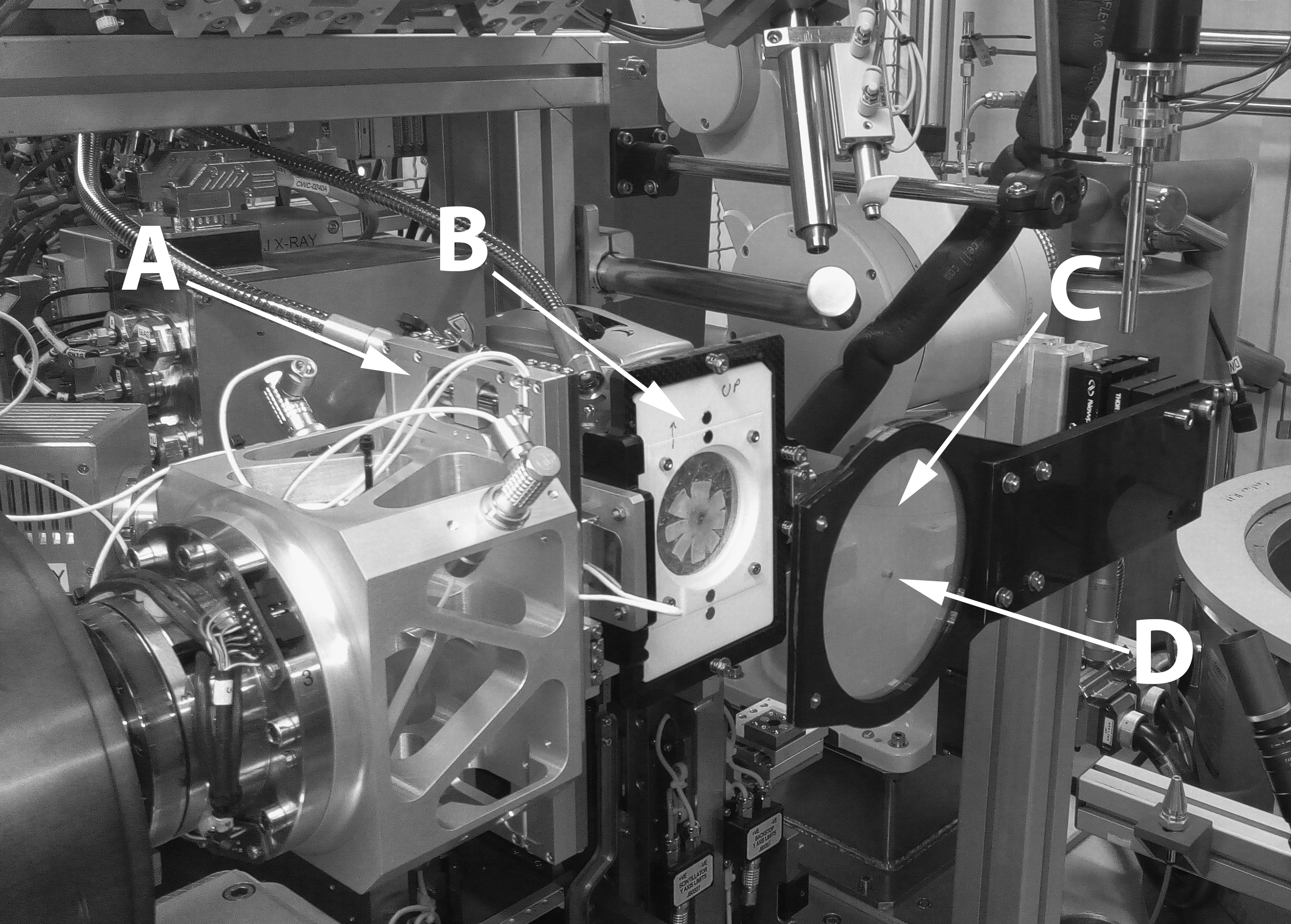
 Figure 2 of
Markov, Mol Vis 2018; 24:818-833.
Figure 2 of
Markov, Mol Vis 2018; 24:818-833.