Figure 1. Calculating the distance between the edge of the optic nerve canal and the tendon insertions of the inferior oblique muscle.
Wide-angle X-ray scattering (WAXS) polar vector plots (plot interval: 0.5 mm) reveal circumferential collagen annulus around
the canal and the oblique uniaxial alignment of the muscle insertion region. The canal edge is denoted by a curved line. Three
individual measurements (line lengths) were performed and a mean taken as the representative value. A: Non-myopic posterior sclera N6. B: Highly myopic specimen HM2. Note the marked increase in line length for the myopic specimen, indicative of the axial lengthening
of the globe.
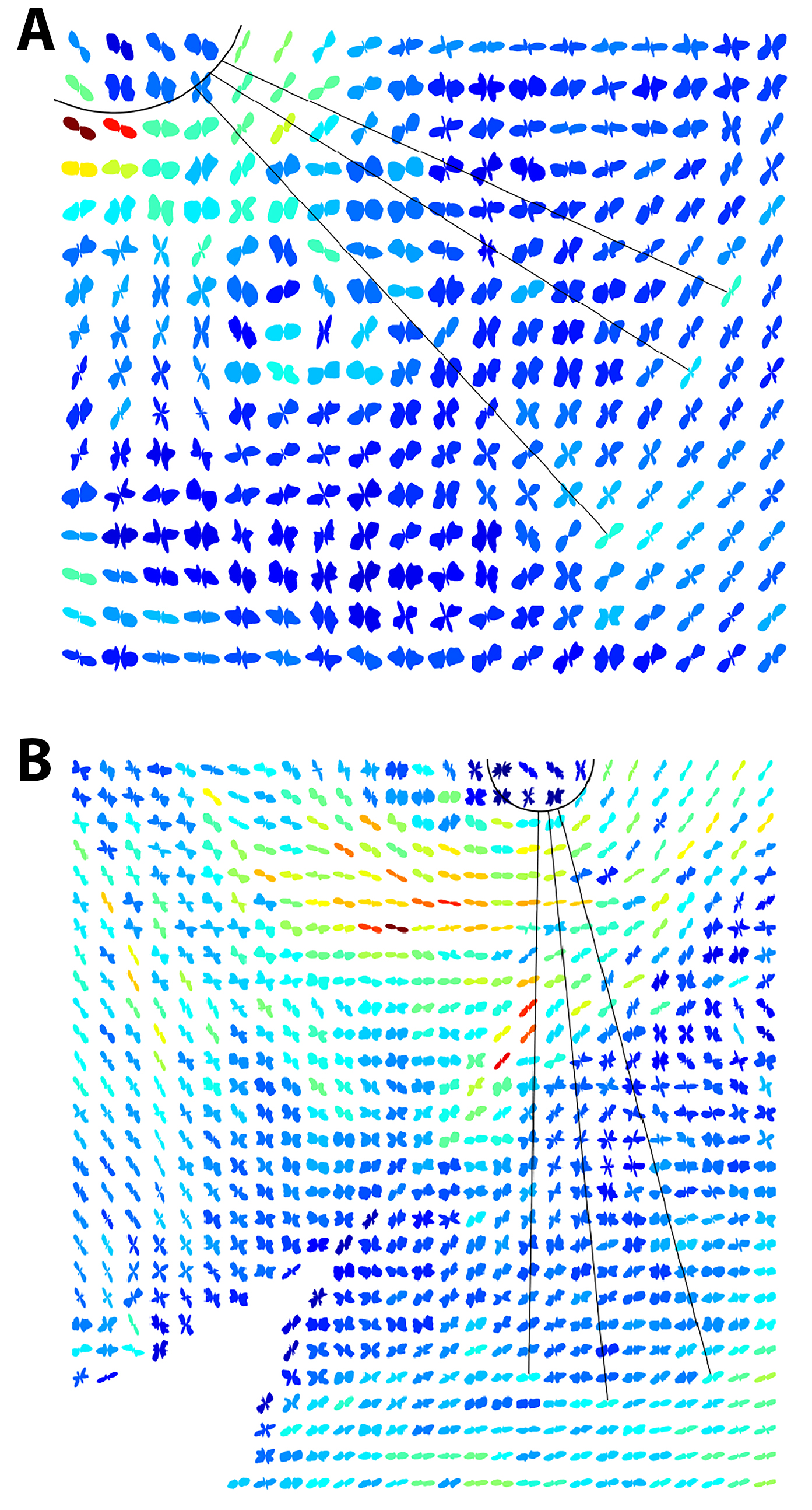
 Figure 1 of
Markov, Mol Vis 2018; 24:818-833.
Figure 1 of
Markov, Mol Vis 2018; 24:818-833.