Figure 2. Presence of iron-free hemoglobin in PEX material and lens epithelium. Sections of (A) pseudoexfoliation (PEX)-affected and (B) unaffected lens capsules were stained with Perls’ Prussian blue and imaged in bright-field at 60X original magnification.
Positive staining would appear blue. Neither the PEX material deposited on the affected capsule (arrows in A) nor lens epithelial cells in the unaffected capsule (B) showed positive staining indicating that the hemoglobin present at these sites is free of iron. Lens epithelial cell nuclei
are counterstained red in panel B.
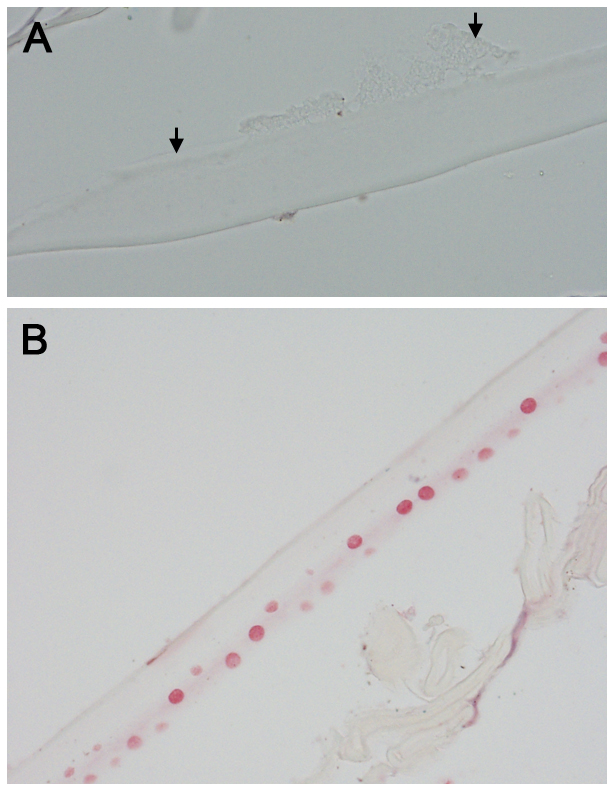
 Figure 2 of
Sharma, Mol Vis 2018; 24:801-817.
Figure 2 of
Sharma, Mol Vis 2018; 24:801-817.