Figure 1. Immunohistochemical labeling of novel proteins identified in PEX material with mass spectrometry in pathological deposits
on PEX-affected lens capsules. Sections of pseudoexfoliation (PEX)-affected (left panels) and unaffected (right panels) lens
capsules from patients with cataract were immunolabeled with the anti-APOA1, anti-APOA4, anti-CRYAA, anti-FGB, anti-HB, anti-PRDX2,
or anti-ZO-1 antibody as indicated. Positive labeling in a section (brown) shows the presence of the indicated protein. Each
labeled protein was detected in pathological material deposited on PEX-affected lens capsules (left panels) and in the lens
epithelium in unaffected lens capsules (right panels). ZO-1, an irrelevant protein, was absent in PEX material deposited on
affected lens capsules proving the specificity of the labeling of the other proteins. ZO-1 protein was detected in the lens
epithelium in unaffected lens capsules (right panel), as expected. 2° Ab, negative control sections hybridized with secondary
antibody without primary antibody hybridization. H&E, sections stained with hematoxylin and eosin. Two parts of the lens capsule
seen in some panels are due to folding of the capsule. Apparent positive immunolabeling of the anterior edge of the control
capsule with the anti-APOA1 antibody is a sectioning artifact (APOA1, right panel). Representative images from independent
experiments on three PEX-affected and four unaffected lens capsules are shown. Images are at 60X original magnification.
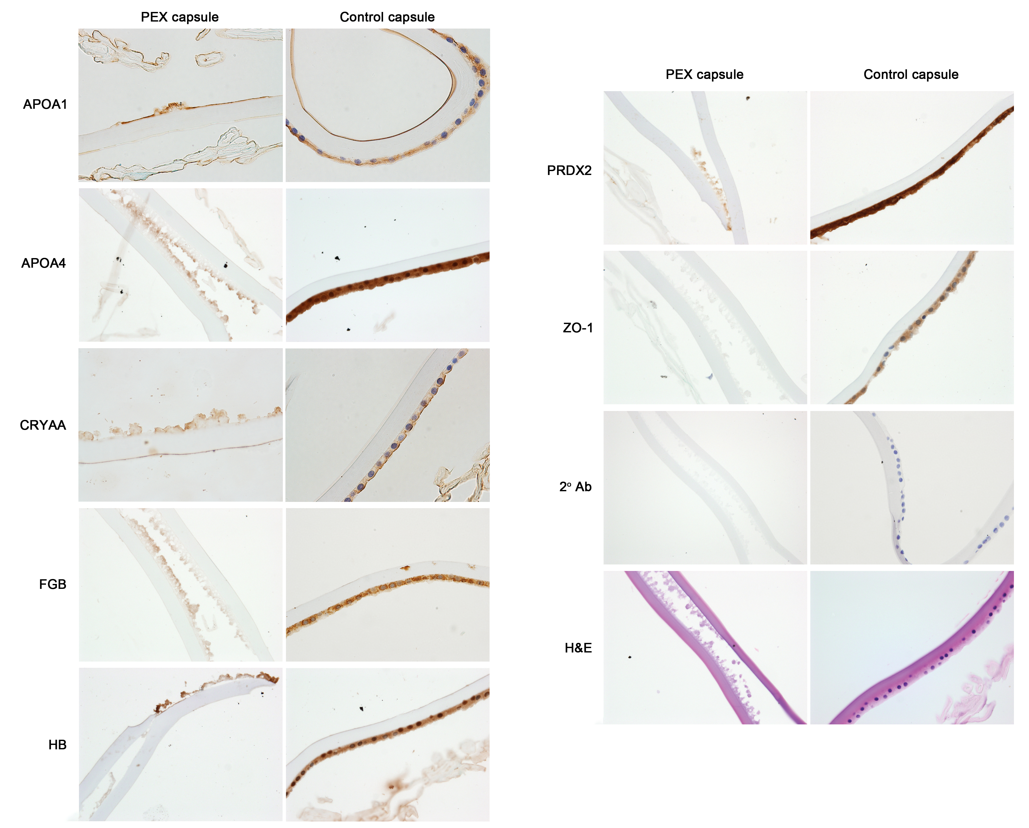
 Figure 1 of
Sharma, Mol Vis 2018; 24:801-817.
Figure 1 of
Sharma, Mol Vis 2018; 24:801-817.