Figure 1. Characterization of human Tenon’s fibroblasts (HTFs). A: HTFs were identified by immunostaining with fibroblast markers, vimentin (green), and nuclei (blue) were labeled with 4’,6-diamidino-2-phenylindole
(DAPI). Scale bar = 100 μm. B: HTFs were immunostained with the epithelial cell marker, cytokeratin, and nuclei (blue) were labeled with DAPI. Scale bar
= 100 μm. C: Lens epithelial cells were immunostained with the epithelial cell marker, cytokeratin, and nuclei (blue) were labeled with
DAPI. Scale bar = 100 μm.
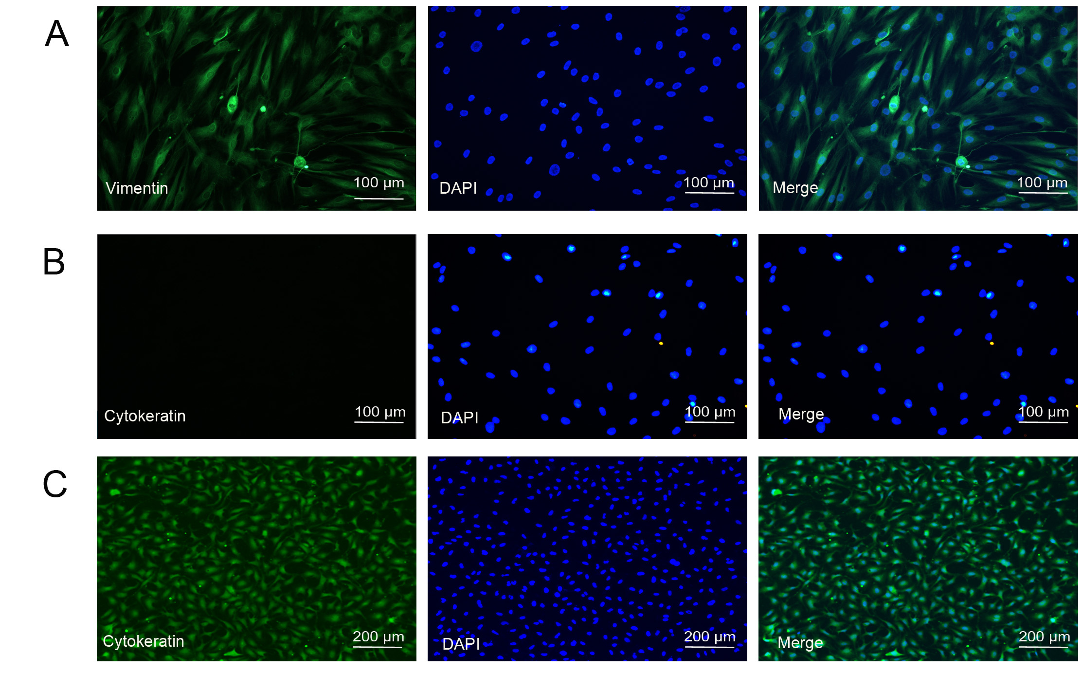
 Figure 1 of
Lin, Mol Vis 2018; 24:789-800.
Figure 1 of
Lin, Mol Vis 2018; 24:789-800.