Figure 5. Examples of segmentations of the peripheral avascular area. The left column contains the unmodified lectin-stained retinal
flat mounts. The middle column contains manual segmentations by one grader, and the right column shows the output of A3ID.
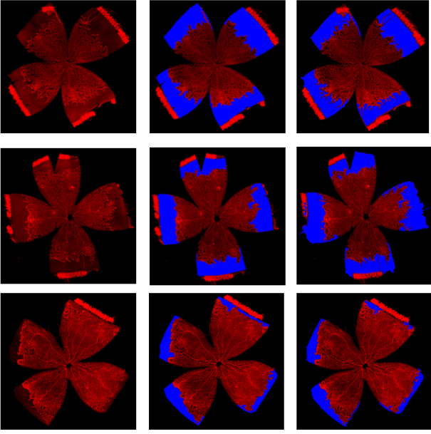
 Figure 5 of
Simmons, Mol Vis 2018; 24:767-777.
Figure 5 of
Simmons, Mol Vis 2018; 24:767-777.