Figure 1. Comparison between rat and mouse retinas in oxygen-induced retinopathy models; a=ciliary body, b=avascular retina, c=vascular
retina, and d=background. There is a central avascular zone in the mouse OIR model and a peripheral avascular zone in the
rat OIR model.
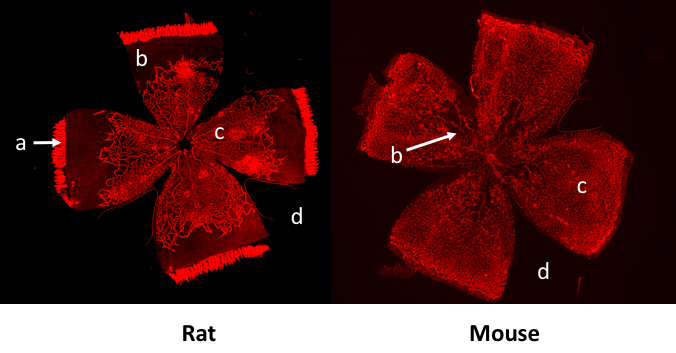
 Figure 1 of
Simmons, Mol Vis 2018; 24:767-777.
Figure 1 of
Simmons, Mol Vis 2018; 24:767-777.