Figure 4. Immunohistochemical staining for TTR. Mass spectrometry revealed upregulation of transthyretin (TTR) which was further characterized
with immunohistochemistry. Staining for TTR revealed that TTR was present in all retinal layers and in the choroid. A strong
reaction for TTR was seen in the retinal vessels regardless of bevacizumab intervention. A: In branch retinal vein occlusion (BRVO) with bevacizumab intervention, a strong reaction for TTR was observed in the choroid.
In the choroid, the reaction for TTR was stronger in BRVO with bevacizumab intervention (A) compared to BRVO without bevacizumab intervention (B). Therefore, the choroid may be the source of increased TTR in BRVO with bevacizumab intervention. Scale bar = 50 µm. Reaction
color: brown. Abbreviations: ILM: inner limiting membrane; NFL: nerve fiber layer; GCL: ganglion cell layer; IPL: inner plexiform
layer; INL: inner nuclear layer; OPL: outer plexiform layer; ONL: outer nuclear layer; PRL: photoreceptor layer. RPE: retinal
pigment epithelium. CHO: choroid.
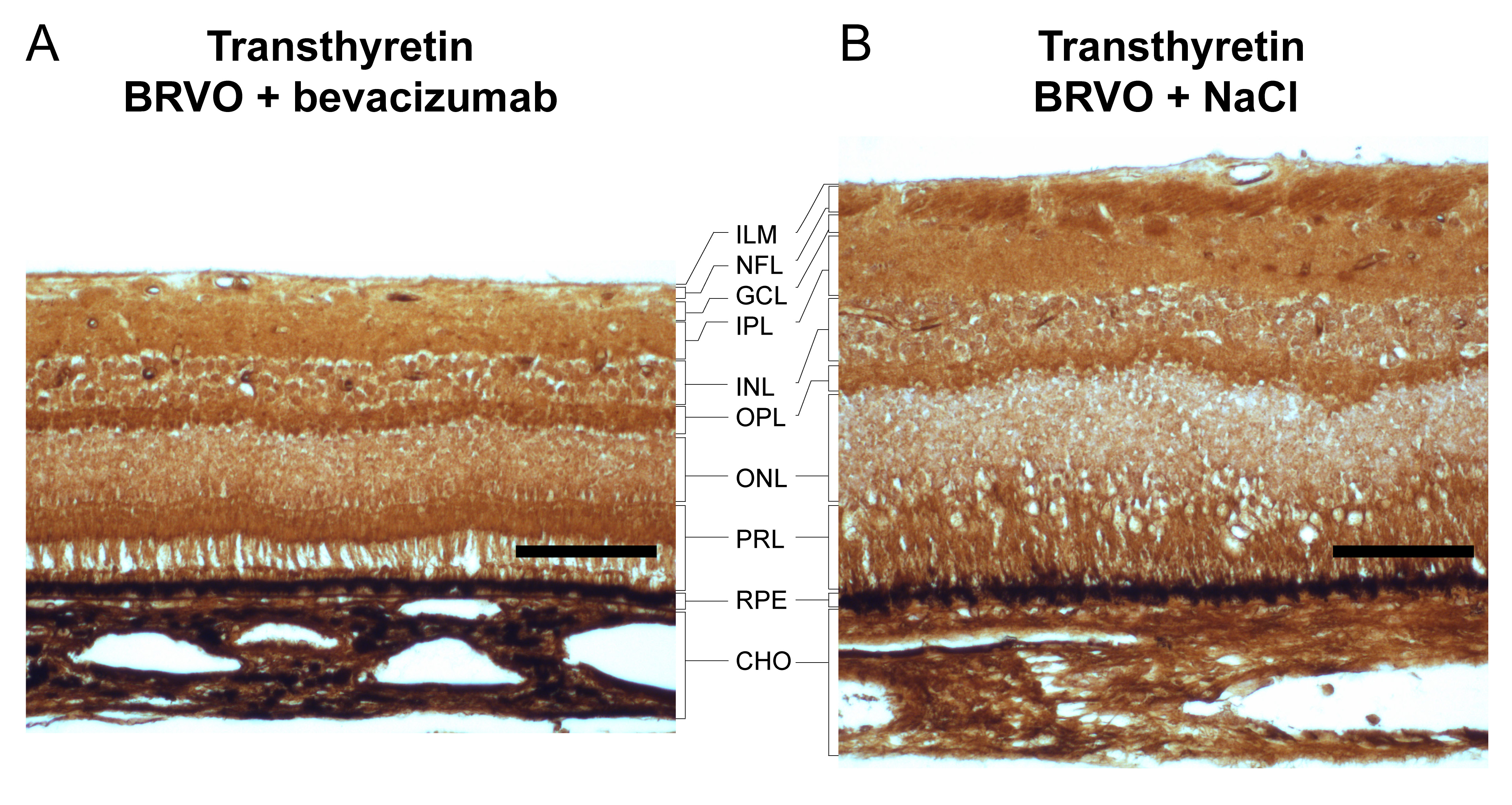
 Figure 4 of
Cehofski, Mol Vis 2018; 24:759-766.
Figure 4 of
Cehofski, Mol Vis 2018; 24:759-766.