Figure 3. Fluorescein angiography was obtained 5 days after BRVO to confirm that no recanalization of the occluded veins had occurred.
Images from the same animal are shown. No passage of fluorescein through the sites of occlusion is observed. A: Fluorescein angiography of branch retinal vein occlusion (BRVO) with bevacizumab intervention. White arrow: site of occlusion.
B: Fluorescein angiography of BRVO without bevacizumab intervention. White arrow: site of occlusion.
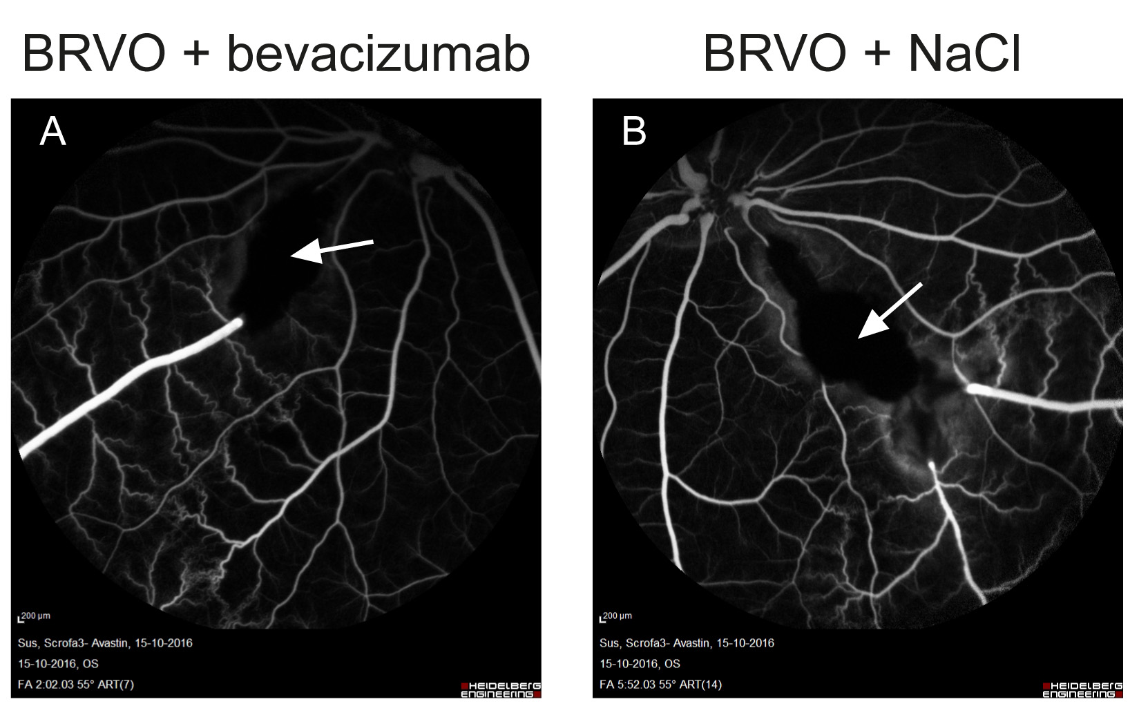
 Figure 3 of
Cehofski, Mol Vis 2018; 24:759-766.
Figure 3 of
Cehofski, Mol Vis 2018; 24:759-766.