Figure 2. Fundus image of BRVO obtained approximately 10 min after BRVO in the inferior retina. Branch retinal vein occlusion (BRVO)
was considered successful when venous dilation and hemorrhages were observed upstream of the site of occlusion as seen in
the image. Black arrow: site of occlusion.
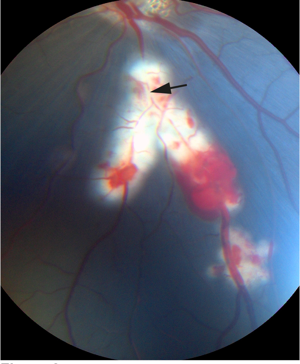
 Figure 2 of
Cehofski, Mol Vis 2018; 24:759-766.
Figure 2 of
Cehofski, Mol Vis 2018; 24:759-766.