Figure 5. The rescue of retinal function in rhodopsin knockout mice with microspheres A: Photopic electroretinography recordings from rho (−/−) mice at 6, 9, and 12 weeks old. The amplitude of the b-waves was measured in glial cell line–derived neurotrophic factor/vitamin
E (GDNF/VitE)-loaded microspheres (MSs), GDNF/VitE/melatonin-loaded MSs, VitE-loaded MSs, and untreated rho (−/−) mice. An averaged electroretinogram (ERG) from a 3-week-old wild-type (wt) mouse and a 3-week-old rho (−/−) mouse is also included. Error bars indicate the standard error of the mean (SEM). B: Comparison of photopic electroretinography recordings from rho (−/−) mice at 6, 9, and 12 weeks after intravitreal injection of microspheres. The amplitude of the b-waves was compared
between the GDNF/VitE-loaded MSs, GDNF/VitE/melatonin-loaded MSs, VitE-loaded MSs, and untreated rho (−/−) mice. Error bars indicate standard error of the mean (SEM); levels of significance: *p<0.05, **p<0.01, ***p<0.001.
Sampling size for each group: 11 animals/group. (graph shows only animals that were included in the analysis).
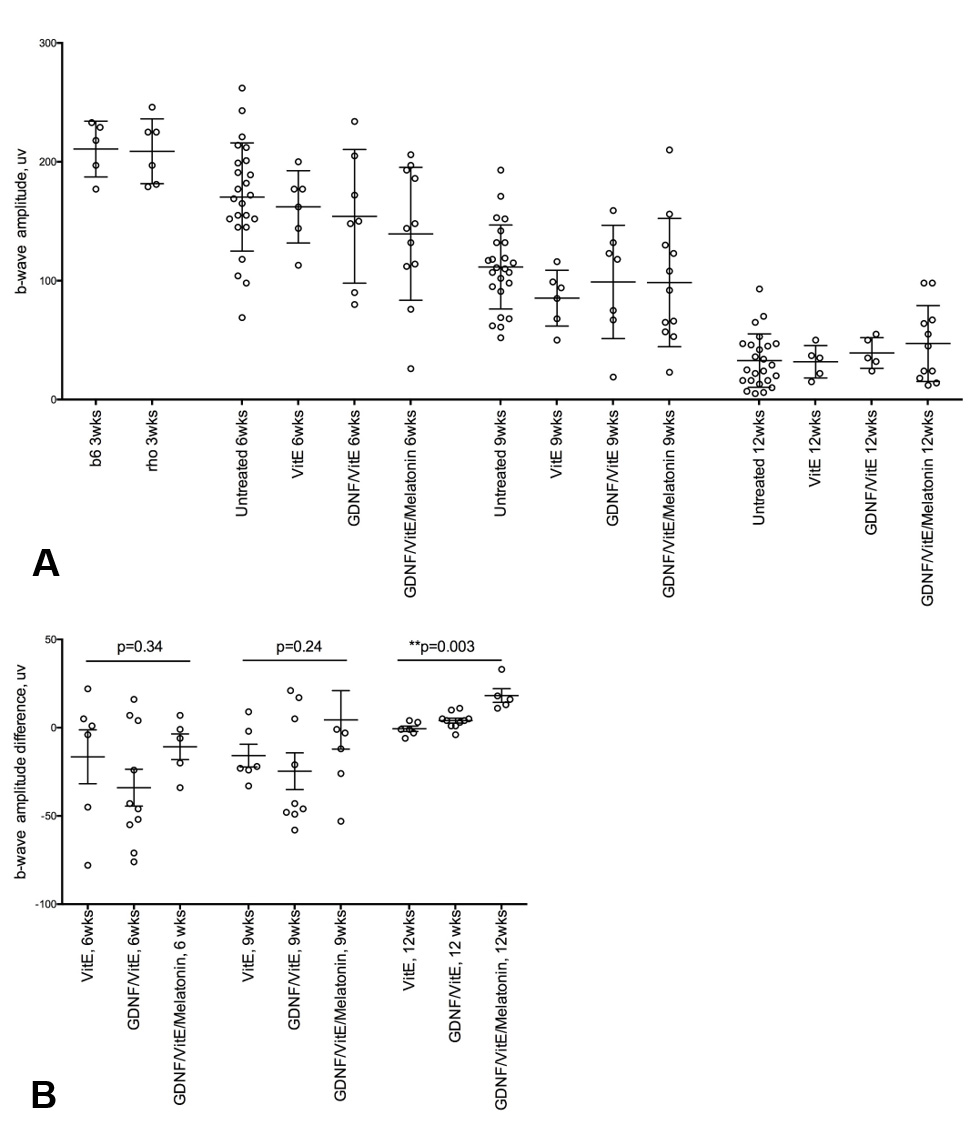
 Figure 5 of
García-Caballero, Mol Vis 2018; 24:733-745.
Figure 5 of
García-Caballero, Mol Vis 2018; 24:733-745.