Figure 4. Immunohistochemical analysis. A: Glial marker expression. The expression of glial markers glial fibrillary acidic protein (GFAP), LIM/homeobox 2 protein
(Lhx2), and glutamine synthetase (GS) was similar across the control and treatment groups. The Müller glia processes are visible
spanning the entire retinal thickness. Scale bar, 50 um. 4′,6-diamidino-2-phenylindole (DAPI; blue) is used for nuclei counterstaining.
B: Expression of the photoreceptor and synaptic markers. The immunohistochemical analysis of the treated and control eyes at
12 weeks following injection of the microspheres demonstrated partial preservation of the photoreceptors. We observed increased
expression of the pan-photoreceptor marker recoverin in both treatment groups, which correlates with increased cell counts
and outer nuclear layer (ONL) thickness. We also confirmed the preservation of outer segments in short-wavelength cones as
detected with S-opsin. The increased expression of synaptic markers vesicle-associated membrane protein (VAMP) and synaptophysin
correlated with the partial photoreceptor rescue in the treatment groups. Scale bar, 50 um. DAPI (blue) is used for nuclei
counterstaining.
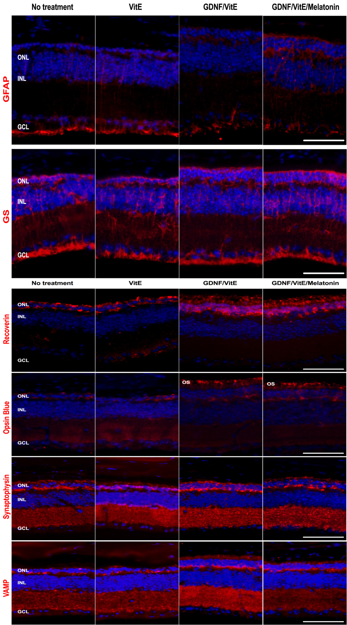
 Figure 4 of
García-Caballero, Mol Vis 2018; 24:733-745.
Figure 4 of
García-Caballero, Mol Vis 2018; 24:733-745.