Figure 5. Brn3a+ RGC counts in radial eye sections. Standardized areas of the ganglion cell layer are shown in sections immunohistochemically
stained for Brn3a (red) prepared from rats intracamerally (IC) injected with TGF-β1+siRhoA for 7 days (A), TGF-β1+siEGFP for 7 days (B), TGF-β1+siRhoA for 35 days (C), TGF-β1+siEGFP for 35 days (D), TGF-β1 for 7 days (E), PBS for 7 days (F), TGF-β1 for 35 days (G), and PBS for 35 days (H), as well as untreated Intact eyes (I). All images are representative of the eight images taken per retina from six different animals (scale bar=50 µm). All images
are nuclear counterstained with 4′,6-diamidino-2-phenylindole (DAPI) (blue). In (J), the mean number of Brn3a+ RGC/mm of retina is presented with counts from the control PBS-treated eyes shown by a dashed line. Asterisks indicate the
significantly lower (p<0.01) RGC counts in the 35 day groups compared to the counts made from their respective 7 day groups
as well as the PBS-treated and Intact control groups.
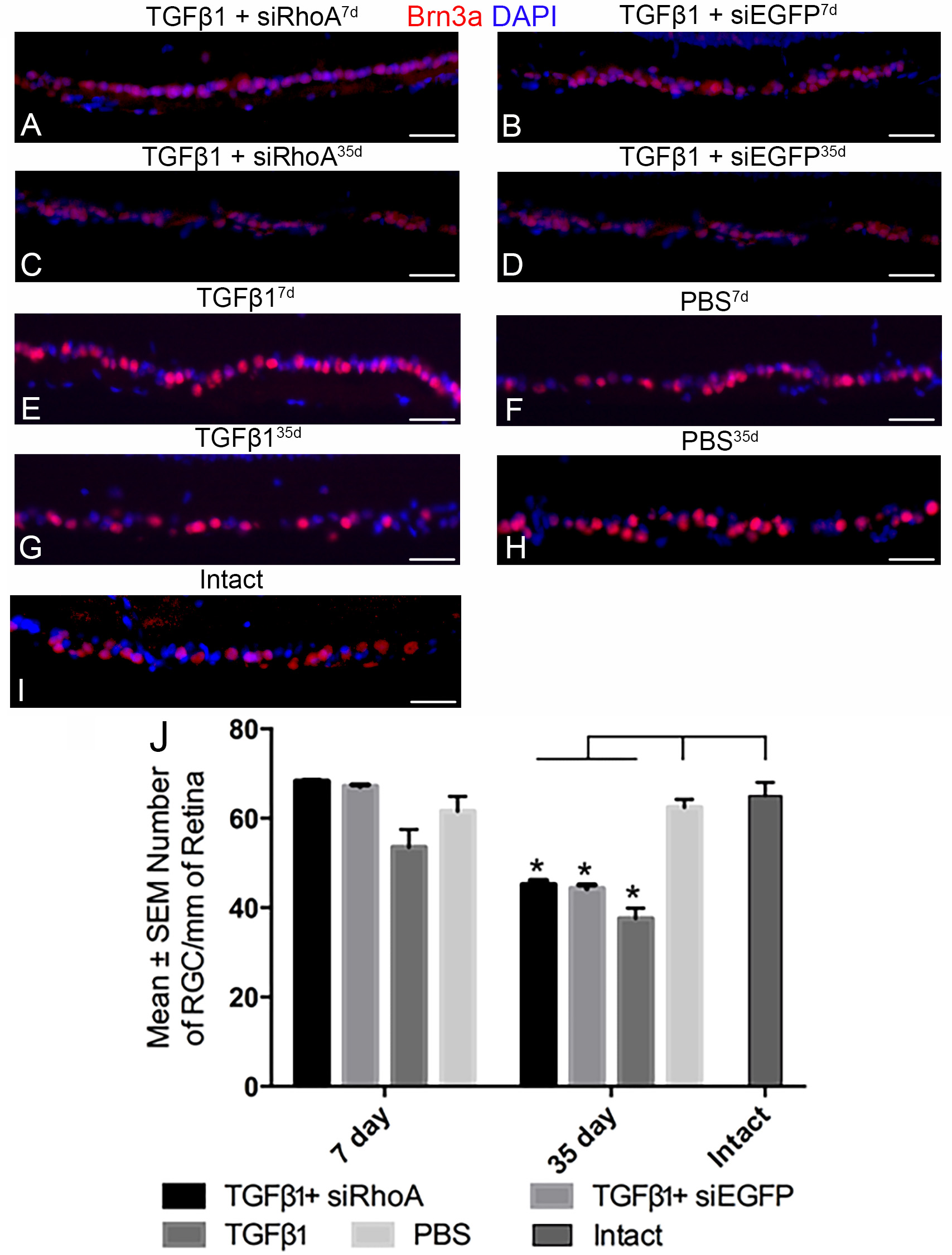
 Figure 5 of
Hill, Mol Vis 2018; 24:712-726.
Figure 5 of
Hill, Mol Vis 2018; 24:712-726.