Figure 3. Percentage of IR fibronectin and laminin pixel counts in the TM of radial eye sections. Images were taken of defined areas
centered on the trabecular meshwork (TM) from radial sections of eyes that were immunohistochemically stained for fibronectin
or laminin (red). Samples were assessed after intracameral (IC) injection with TGF-β1+siRhoA for 7 days (A and E), TGF-β1+siEGFP for 7 days (B and F), TGF-β1+siRhoA for 35 days (C and G), TGF-β1+siEGFP for 35 days (D and H), TGF-β1 for 7 days (I and N), PBS for 7 days (J and O), TGF-β1 for 35 days (K and P), and PBS for 35 days (L and Q), as well as untreated Intact eyes (M and R). All images are representative of eight images taken per retina from each treatment group (scale bar=50 µm). All sections
were nuclear counterstained with 4′,6-diamidino-2-phenylindole (DAPI) (blue). The percentage immunoreactivity (IR) pixel counts in the TM are presented for fibronectin (S) and laminin (T) from the groups defined in A–R. Asterisks indicate the statistically significant differences at p<0.01 between the same treatments at 7 days and the 35
days, whereas the black lines indicate statistically significant differences at p<0.05 between groups at 35 days.
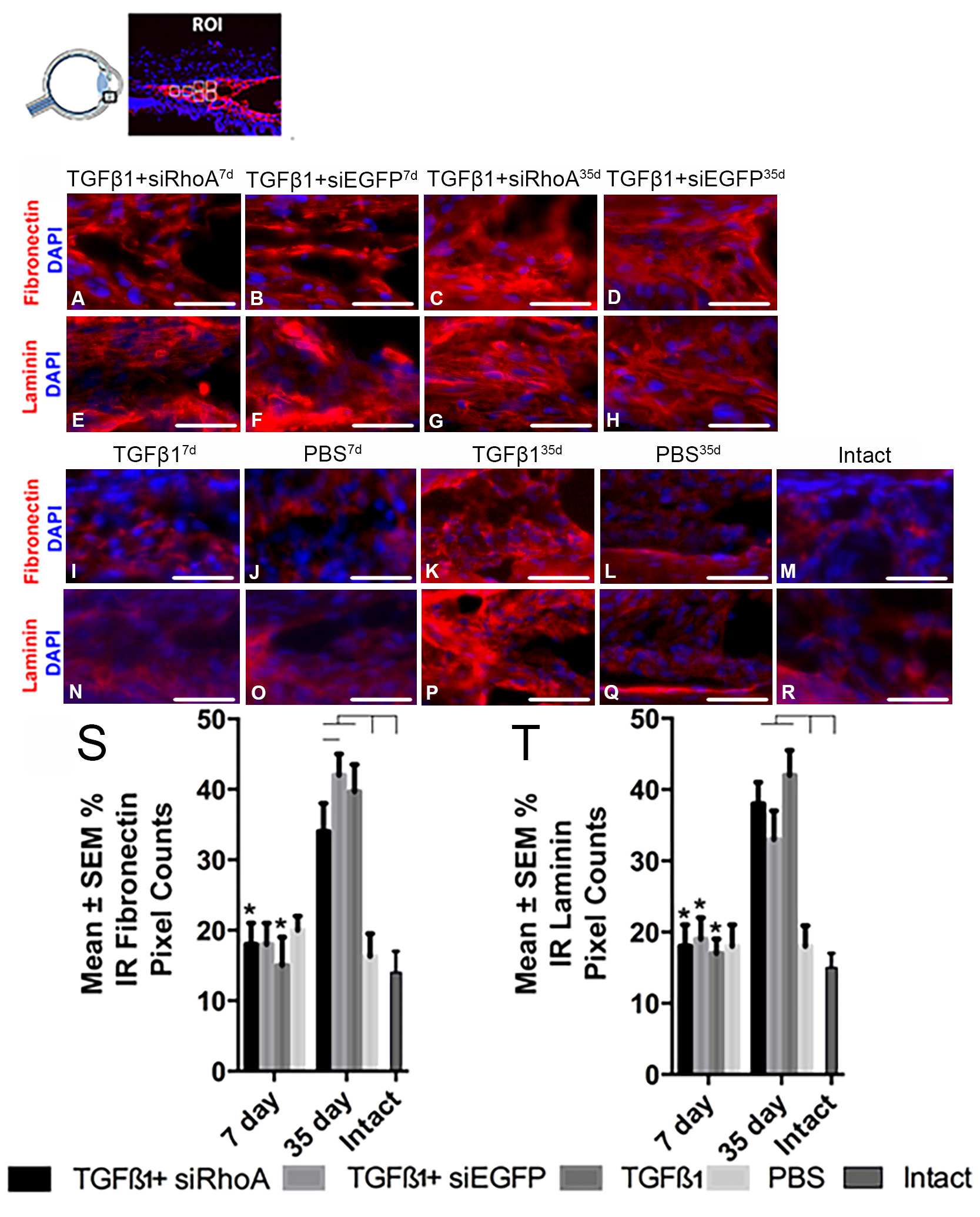
 Figure 3 of
Hill, Mol Vis 2018; 24:712-726.
Figure 3 of
Hill, Mol Vis 2018; 24:712-726.